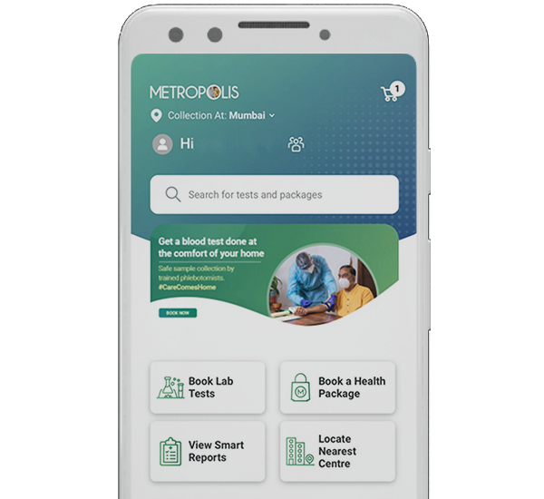
 Home Visit
 Upload


Latest Blogs

Crohn's Disease: Symptoms, Causes, Treatment Options & Types
What is Crohn’s Disease? Crohn's disease is a chronic inflammatory condition that affects the digestive system, especially the small intestine and colon. It is a type of inflammatory bowel disease (IBD) that affects various areas of the digestive system and produces swelling, irritation, and inflammation. What are the Types of Crohn’s Disease? Crohn’s disease can affect different parts of the digestive tract, leading to several distinct types. The most common Crohn's disease types are: Ileocolitis: The most common form, affecting the ileum (last section of the small intestine) and colon (a portion of large intestine) Ileitis: This Crohn's disease type affects only the ileum Gastroduodenal Crohn's Disease: Affects the stomach and the beginning of the small intestine (duodenum) Jejunoileitis: Characterised by patchy areas of inflammation in the upper half of the small intestine (jejunum) Crohn's Colitis: Affects only the colon, similar to ulcerative colitis How Common is Crohn’s Disease? Crohn’s disease is relatively rare, but its occurrence is rising globally. In India, the disease is less common than in Western countries but is becoming more prevalent due to lifestyle shifts and urbanization. Current estimates indicate that about 1 in 10,000 people in India are affected by Crohn’s disease. Increased awareness and better diagnostic methods are leading to more cases being identified. However, it remains underdiagnosed, particularly in rural areas. What are the Symptoms of Crohn’s Disease? Crohn’s disease symptoms vary widely and can range from mild to severe. Common symptoms include: Persistent diarrhoea Abdominal pain and cramping Fatigue Weight loss Blood in stool Fever Reduced appetite Mouth sores Since Crohn's disease symptoms overlap with other gastrointestinal conditions, it often requires a thorough evaluation for accurate diagnosis. The intensity of these symptoms can fluctuate, with periods of remission followed by flare-ups. What are the Complications of Crohn’s Disease? If left untreated, Crohn’s disease can lead to complications such as strictures (narrowed sections of the intestines), fistulas (abnormal connections between the intestines and other organs), and abscesses (pockets of infection). Additionally, Crohn’s disease complications may extend beyond the digestive tract, leading to issues such as arthritis, skin conditions, and eye inflammation. Long-term inflammation can also increase the risk of colon cancer, making regular screenings essential for patients. What Causes Crohn’s Disease? The exact causes of Crohn’s disease are not fully understood, but a combination of factors is believed to contribute to the condition. These factors include genetics, immune system malfunctions, and environmental triggers. For instance, if the immune system mistakenly attacks the digestive tract, it can lead to chronic inflammation. Genetics also play a role, as people with a family history of Crohn’s disease are at higher risk. Environmental factors, such as a western diet high in processed foods, may also contribute to the disease's onset. What are the Risk Factors of Crohn’s Disease? Several risk factors increase the likelihood of developing Crohn’s disease. Family history is one of the most significant risk factors, with those having a close relative with the disease being more susceptible. Smoking is another critical risk factor, as it not only increases the risk of Crohn's disease but also worsens its severity. Age is also a factor, with most cases being diagnosed before the age of 30. Additionally, residing in urban areas or developed countries may expose individuals to environmental triggers that heighten the risk of developing Crohn’s disease. How is Crohn’s disease diagnosed? Lab Tests When diagnosing Crohn's disease, lab tests are often the first step after observing Crohn's disease symptoms such as persistent diarrhoea, abdominal pain, and weight loss. Blood tests are commonly used to check for anaemia, which can occur due to chronic blood loss, and to assess inflammation levels. Stool tests may also be performed to rule out infections and detect blood or inflammatory markers in the stool. These tests help doctors differentiate Crohn's disease from other conditions and guide the next steps in Crohn's disease treatment. Imaging Procedures Imaging procedures play a crucial role in diagnosing Crohn’s disease by providing visual evidence of inflammation and other abnormalities in the digestive tract. Common imaging techniques include CT scans and MRI scans. CT enterography and MR enterography are specialised versions of these scans that focus on the small intestine, which is where Crohn's disease frequently manifests. These treatments aid in the diagnosis of thickened bowel walls, fistulas, abscesses, and other complications related with the cause of Crohn's disease. Ultrasounds may also be used, especially in children and pregnant women, as a less invasive method. Imaging can distinguish between different Crohn’s disease types by showing the extent and location of inflammation in the intestines, aiding in treatment planning. Endoscopy Endoscopy is crucial in the diagnosis of Crohn's disease. This method uses a flexible tube equipped with a camera to provide direct vision of the digestive tract. Colonoscopy and upper endoscopy are the two primary forms of endoscopy that are used. A colonoscopy examines the colon and the end of the small intestine, making it highly effective for detecting inflammation, ulcers, and strictures in these areas. Upper endoscopy, on the other hand, explores the oesophagus, stomach, and upper part of the small intestine. Both procedures enable the collection of biopsies, which are crucial for diagnosing Crohn’s disease by identifying the specific inflammation patterns typical of the disease. Capsule endoscopy is an alternative way to examine the small intestine, especially when other methods have not provided clear results. In this procedure, the patient swallows a small camera. These endoscopic procedures are crucial for differentiating the symptoms and types of Crohn's disease from other inflammatory bowel diseases, which in developing tailored treatment plans for individuals with Crohn's disease. How is Crohn’s disease treated? Medications Medications are a key element in managing Crohn's disease, primarily targeting inflammation reduction and symptom control. Anti-inflammatory drugs, such as aminosalicylates, are often used for mild to moderate cases. Corticosteroids, which are stronger anti-inflammatory drugs, are prescribed for more severe flare-ups, but long-term use is avoided due to potential side effects. Immune system suppressors, like azathioprine or biologics such as infliximab, are used to target specific aspects of the immune response that contribute to Crohn's disease causes. Antibiotics may also be used to treat complications like abscesses and fistulas. Nutrition Diet and nutrition play a vital role in managing Crohn's disease, although they are not direct treatments for the condition. A low-residue diet, which reduces fibre intake, is commonly recommended during flare-ups to minimize irritation of the inflamed digestive tract. In cases where malnutrition is a concern, supplements and special diets, such as enteral nutrition (liquid diets), may be prescribed to ensure adequate nutrient intake. Maintaining a balanced diet and avoiding trigger foods can greatly support the treatment of Crohn's disease, although the most effective strategies may vary from one individual to another. Regular consultations with a dietitian can help in creating a tailored nutrition plan that supports overall health and reduces the impact of Crohn's disease causes. Surgery Surgery becomes necessary for Crohn's disease treatment when medications and dietary changes are ineffective or when complications develop. Approximately 70% of individuals with Crohn's disease will ultimately need surgery. Surgical procedures may involve removing a damaged section of the digestive tract, draining abscesses, or repairing fistulas. In some cases, a procedure known as strictureplasty is performed to widen a narrowed area of the intestine without removing any part of it. Surgery can provide significant relief from Crohn's disease symptoms, but it is not a cure, as the disease often recurs in other parts of the digestive system. Post-surgery, medications and lifestyle changes remain essential to prevent relapse. For many patients, surgery can improve their quality of life when combined with ongoing Crohn's disease treatment plans. How can I prevent Crohn’s disease? Preventing Crohn's disease is challenging because its exact causes are still not fully understood. However, certain lifestyle modifications may help reduce the risk of developing the disease or prevent flare-ups for those already diagnosed. Healthy Diet: There's no specific diet to prevent Crohn's disease, maintaining a balanced diet rich in fruits, vegetables, and whole grains may support gut health. Avoiding processed foods and excessive fats may also reduce inflammation. Quit Smoking: Smoking is a significant risk factor for Crohn's disease. Quitting can not only lower your risk but also improve outcomes for those already diagnosed. Stress Management: High stress levels can trigger flare-ups. Engaging in stress-reducing activities such as yoga, meditation, and regular exercise can help manage Crohn's disease symptoms. Regular Medical Check-ups: If you have a family history of Crohn’s disease, regular check-ups can help with early detection and management. While these steps may not guarantee prevention, they can play a role in reducing the impact of Crohn's disease causes and improving overall well-being. Is there a cure for Crohn’s disease? There is currently no cure for Crohn's disease. However, treatments like medications, diet changes, and occasionally surgery can help control symptoms, decrease inflammation, and enhance quality of life. What is the outlook for people with Crohn’s disease? The outlook can differ; with appropriate treatment and lifestyle changes, many individuals with Crohn's disease can lead healthy lives, although flare-ups and complications may still occur. What is the life expectancy of a person with Crohn’s disease? Most people with Crohn's disease have a normal life expectancy, especially with effective treatment and regular medical care to manage symptoms and complications. How does Crohn’s disease affect pregnancy? Crohn's disease can influence pregnancy, but many women with the condition can still have healthy pregnancies with careful management. Flare-ups are more likely if the disease is active during conception. When to see a doctor? See a doctor if you experience persistent gastrointestinal symptoms such as severe abdominal pain, diarrhoea, weight loss, or blood in the stool, as these may indicate Crohn's disease. Regular check-ups are essential for managing the condition. Conclusion Living with Crohn's disease can be challenging, but with early diagnosis and effective management can help individuals lead fulfilling lives. For accurate testing and diagnosis, trust Metropolis Labs. Offering advanced services, expert consultations, and home sample collections, Metropolis Labs ensures you receive the precise care you need.

Angioplasty: Surgery, Procedure, Types, Benefits, Complications and Recovery
What is an Angioplasty? Angioplasty is a medical procedure that helps to restore blood flow through arteries that are narrowed or blocked. This procedure often performed to treat heart problems caused by plaque buildup in the coronary arteries, which can reduce blood flow to the heart. Angioplasty surgery usually involves inserting a small balloon into a blocked artery and inflating it to widen the artery. A stent may also be added to keep the artery open. This minimally invasive procedure helps to increase blood flow and reduces the risk of heart attacks. Who Needs to Have Angioplasty? Angioplasty is recommended for individuals with coronary artery disease (CAD), where the arteries supplying blood to the heart become narrowed due to plaque buildup. People with lifestyle choices like poor diets, smoking, and insufficient physical activity have contributed to an increase in cases of coronary artery disease (CAD). Patients experiencing chest pain (angina), shortness of breath, or those who have had a heart attack may be candidates for angioplasty surgery. It is also suggested for those who have not responded well to medication or lifestyle changes aimed at reducing heart disease risks. What Does Angioplasty Treat? Angioplasty is used to treat blocked or narrowed coronary arteries, which are responsible for supplying oxygen-rich blood to the heart. This condition is often associated with heart disease, which is a leading cause of death. Angioplasty is an effective procedure in relieving angina symptoms, reducing the risk of heart attacks, and enhancing the overall quality of life for individuals with heart disease. Additionally, it can be used to address peripheral artery disease (PAD), which involves the narrowing of arteries in areas such as the legs. How Common is Angioplasty? Angioplasty surgery has become increasingly common in India, especially in urban areas where advanced medical resources are readily available. With the rising incidence of heart disease in the country, angioplasty is a frequently performed procedure. According to reports, India has over a million angioplasty procedures each year, making it one of the most common treatments for coronary artery disease. The easy accessibility and availability of this procedure have made it a preferred option for many patients. What Happens Before Angioplasty? Before undergoing angioplasty, a series of evaluations and tests are conducted to make sure the patient is a suitable candidate for the procedure. This typically includes blood tests, an electrocardiogram (ECG), and imaging tests like coronary angiography to visualise the blocked arteries. Patients are usually advised to fast for a few hours before the angioplasty procedure and may be asked to stop certain medications, particularly blood thinners. It’s also important for the patient to discuss any existing medical conditions with their doctor to avoid any complications during the procedure. What Happens During an Angioplasty? Angioplasty is a minimally invasive procedure primarily designed to treat narrowed or blocked arteries, most commonly in the heart. Angioplasty procedure aims to restore blood flow through the arteries, preventing complications like heart attacks or severe chest pain (angina). This procedure usually takes 1 to 2 hours and is conducted in a catheterization lab (cath lab) by an interventional cardiologist. Before the angioplasty, the patient receives a mild sedative to promote relaxation, while still remaining responsive during the procedure. A local anesthetic is applied to numb the area where the catheter will be inserted, typically in the groin or wrist. Once the area is numb, the doctor makes a small incision and inserts a thin, flexible tube called a catheter into the artery. The catheter is gently navigated through the blood vessels to reach the blockage in the coronary artery. During this process, the doctor uses real-time X-ray images, known as fluoroscopy, to see the exact position of the catheter. This ensures precise navigation through the arteries. Once the catheter reaches the blockage, a small balloon at the tip of the catheter is inflated. This balloon presses the plaque against the artery walls, widening the artery and improving blood flow. This part of the angioplasty surgery is crucial, as it helps to alleviate symptoms and reduces the risk of future heart-related problems. In many cases, a stent is inserted into the artery during the angioplasty procedure. A stent is a small, mesh-like metal tube that acts as a support to keep the artery open. The stent is attached to the balloon catheter and expands when the balloon is inflated. Once the stent is positioned, the balloon is deflated and removed, leaving the stent permanently in the artery to prevent it from narrowing again. Throughout the angioplasty, the patient’s vital signs are closely monitored, and any discomfort is addressed immediately. Most patients describe sensations of pressure or mild discomfort during the balloon inflation, but they generally do not feel intense pain. Once the procedure is complete, the catheter is carefully removed, and the incision site is closed using manual pressure, a closure device, or stitches. What Happens After Angioplasty? After angioplasty, patients are transferred to a recovery area where they are monitored for several hours. The recovery period may vary depending on the hospital and patient's overall health status. Generally, most patients are discharged within 24 to 48 hours if no complications arise during the procedure. It’s essential to follow doctor’s advice regarding medications, particularly blood thinners, and attend follow-up appointments to ensure the artery stays open. Additionally, lifestyle changes such as a heart-healthy diet, regular exercise, and quitting smoking is advised to sustain the benefits of the angioplasty. What are the Benefits of Angioplasty After a Heart Attack? Angioplasty is particularly beneficial following a heart attack, as it restores blood flow to the heart and reduces damage to the heart muscle. In India, where the incidence of heart attacks are becoming more frequent, a prompt angioplasty can greatly enhance survival rates and lower the risk of future heart issues. The procedure also helps in relieving symptoms like chest pain and shortness of breath, allowing patients to return to their daily activities more quickly. Additionally, angioplasty reduces the need for more invasive surgeries like coronary artery bypass grafting (CABG). What are the Risks or Complications of Angioplasty? While angioplasty is usually safe, it does come with some risks, like any other medical procedure. In India, where heart disease is common, it's crucial to be informed about potential complications, including bleeding at the catheter insertion site, blood vessel damage, or allergic reactions to the dye used during the procedure. In rare cases, serious issues like heart attacks, strokes, or the need for emergency surgery can occur. Another concern is restenosis, which is re-narrowing of the artery, but using drug-eluting stents has made this less common. It is important for patients to discuss these potential risks with their doctor before undergoing the procedure. What is the Recovery Time After Angioplasty? Recovery time after angioplasty varies, but most patients in India can resume their usual activities within a week. It’s important to avoid heavy lifting or strenuous workouts for several weeks to ensure proper healing of the artery. Patients are generally advised to make important lifestyle changes to prevent additional heart problems. This includes eating a healthy diet, regular exercise, and quit smoking. Attending follow-up appointments and taking prescribed medications as instructed are also vital for a successful recovery. When to See a Doctor? It’s important to see a doctor if you experience symptoms like chest pain, shortness of breath, or any unusual discomfort after an angioplasty procedure. Timely medical attention can prevent complications and ensure the success of the treatment. Patients should contact their doctor if they observe any signs of infection at the catheter insertion site, including redness, swelling, or discharge. Regular check-ups are essential to monitor heart health and address any concerns early on. Conclusion Angioplasty is a life-saving procedure that has become more prevalent in India due to the growing incidence of heart disease. By following medical guidance and adopting healthy lifestyle habits, individuals can improve the long-term success of angioplasty and enhance their overall quality of life. Metropolis Labs is a leading choice across India for accurate diagnostics and superior care. Equipped with state-of-the-art testing and a team of highly trained professionals, Metropolis Labs provides comprehensive health check-ups, including cardiac profiles essential before and after angioplasty. Trust Metropolis for precise and timely results, enabling you and your doctor to make well-informed decisions regarding your heart health.

आयरन युक्त खाद्य पदार्थ: अपने स्वास्थ्य को प्राकृतिक रूप से बेहतर बनाएँ
आयरन युक्त खाद्य पदार्थ क्या हैं? आयरन युक्त खाद्य पदार्थ वे होते हैं जिनमें आयरन की मात्रा अधिक होती है। यह महत्वपूर्ण खनिज हमारे शरीर में वृद्धि, विकास और स्वस्थ रक्त कोशिकाओं के निर्माण के लिए आवश्यक है। जब लोग आयरन-युक्त भोजन के बारे में सोचते हैं, तो अक्सर एक स्वादिष्ट स्टेक या पालक का भारी हिस्सा उनके दिमाग में आता है। लेकिन सच्चाई यह है कि आयरनसे भरपूर कई अन्य विकल्प उपलब्ध हैं। मछली से लेकर अंडे, नट्स से लेकर दालें और यहां तक कि कई तरह के फल और सब्जियां ये सभी आयरन युक्त खाद्य स्रोत हैं। आपको प्रति दिन कितने आयरन की आवश्यकता है? आपको प्रतिदिन कितने आयरन की आवश्यकता होती है, यह आपकी उम्र, लिंग और स्वास्थ्य स्थिति पर निर्भर करता है। 19 वर्ष और उससे अधिक उम्र के वयस्क पुरुषों के लिए, अनुशंसित दैनिक मात्रा आमतौर पर 8 मिलीग्राम (mg) होती है। 19-50 वर्ष की वयस्क महिलाओं को मासिक धर्म के रक्तस्राव के कारण प्रतिदिन लगभग 18 mg की आवश्यकता होती है। रजोनिवृत्ति के बाद, अनुशंसित मात्रा घटकर 8 mg प्रतिदिन हो जाती है। गर्भवती महिलाओं को भ्रूण के विकास और रक्त की मात्रा में वृद्धि का समर्थन करने के लिए प्रति दिन 27 mg तक की अधिक आवश्यकता होती है। आयरन शरीर में ऑक्सीजन के परिवहन और ऊर्जा उत्पादन में महत्वपूर्ण भूमिका निभाता है, और आयरन की कमी, जो थकान, कमजोरी और संज्ञानात्मक कार्य में कमी का कारण बन सकती है, इस बात पर जोर देती है कि आयरन युक्त खाद्य पदार्थों का सेवन कितना महत्वपूर्ण है। हेम और नॉन-हेम आयरन में क्या अंतर है? जब आप आयरन युक्त खाद्य पदार्थ खाते हैं, तो यह मुख्य रूप से आपकी छोटी आंत के माध्यम से अवशोषित होता है। भोजन से प्राप्त होने वाले आयरन के दो प्रकार हैं - हेम और नॉन-हेम। हेम आयरन लाल मांस, मछली और मुर्गी जैसे पशु खाद्य पदार्थों में पाया जाता है, जिनमें पहले से ही हीमोग्लोबिन होता है। हमारे शरीर के लिए इस प्रकार के आयरन को अवशोषित करना आसान होता है। नॉन-हेम आयरन ज्यादातर पौधों के स्रोतों या पालक, बीन्स, समृद्ध अनाज और सेरेल्स जैसे फोर्टिफाइड खाद्य पदार्थों से आता है। हालांकि, कुछ नॉन-हेम आयरन मांस, मुर्गी और सीफूड में भी मौजूद होता है, क्योंकि ये जानवर पौधों से प्राप्त खाद्य पदार्थों का सेवन करते हैं। आयरन-युक्त खाद्य पदार्थ स्वास्थ्य को बनाए रखने के लिए आयरन युक्त खाद्य पदार्थ खाना आवश्यक है, क्योंकि आयरन शरीर में ऑक्सीजन के परिवहन में महत्वपूर्ण भूमिका निभाता है। अपने आहार में आयरन युक्त खाद्य पदार्थों को शामिल करने से इस आवश्यक खनिज का पर्याप्त स्तर सुनिश्चित होता है, जो ऊर्जा उत्पादन और समग्र स्वास्थ्य का समर्थन करता है। इन खाद्य पदार्थों का विटामिन C के स्रोत के संयोजन से आयरन अवशोषण में सुधार हो सकता है। अपने आहार में आयरन युक्त विभिन्न खाद्य पदार्थों को शामिल करने से आयरन की कमी को रोकने और समग्र जीवन शक्ति को बढ़ावा देने में मदद मिल सकती है। आयरन से भरपूर मांस: कुछ मांस जिनमें आयरन की मात्रा अधिक होती है, उनमें लिवर, बीफ, चिकन, पोर्क, हिरन और भेड़ का मांस शामिल हैं। आयरन से भरपूर सीफूड: समुद्री भोजन में ऑयस्टर, मसल्स, श्रिम्प, क्लैम, सार्डिन, मैकेरल, टूना और स्कैलप्स शामिल हैं। आयरन से भरपूर सब्जियां: आयरन से भरपूर सब्जियों में पालक, केल, कोलार्ड ग्रीन्स, बीट ग्रीन्स, चार्ड, स्वीट पोटैटो, ब्रोकली और स्ट्रिंग बीन्स शामिल हैं। आयरन से भरपूर फल: आयरन से भरपूर फलों में स्ट्रॉबेरी, तरबूज, अंजीर, खजूर, किशमिश, ड्रॉयड अपरिकोट्स और प्लम शामिल हैं। अन्य खाद्य पदार्थ जिनमें आयरन की मात्रा अधिक होती है अन्य आयरन युक्त खाद्य पदार्थों में अंडे, बीन्स, दाल, आयरन-फोर्टिफाइड सेरेल्स, ब्रेड और पास्ता, शीरा, मटर और मेपल सिरप शामिल हैं। अब जब हम विभिन्न आयरन युक्त खाद्य पदार्थों के बारे में जानते हैं, तो आइए उन अलग-अलग खाद्य पदार्थों पर ध्यान दें जिनमें आयरन की मात्रा अधिक होती है: पालक: आयरन की मात्रा की बात करें तो पालक एक सुपरफूड है। सिर्फ एक कप पके हुए पालक में 6 mg से ज़्यादा आयरन होता है। शेलफिश: क्लैम और ऑयस्टर जैसी शेलफिश न सिर्फ स्वादिष्ट होती हैं बल्कि हेम आयरन से भी भरपूर होती हैं, जिसे शरीर आसानी से अवशोषित कर सकता है। टोफू: शाकाहारियों और वेगन लोगों के लिए आदर्श, टोफू नॉन-हेम आयरन का एक बेहतरीन स्रोत है। आधे कप में लगभग 3.4 mg आयरन होता है। पोल्ट्री, रेड मीट और मछली: चिकन लिवर की एक सर्विंग में लगभग 13 mg आयरन होता है, जो सूची में सबसे ऊपर है। बीफ स्टेक और ग्राउंड मीट भी अच्छी मात्रा में आयरन प्रदान करते हैं। टूना और सार्डिन जैसी मछलियां भी पीछे नहीं हैं। व्होल ग्रेन्स: क्विनोआ या फोर्टिफाइड सेरेल्स जैसे व्होल ग्रेन्स आपके दैनिक आयरन सेवन में महत्वपूर्ण योगदान दे सकते हैं। डार्क चॉकलेट: क्या आप जानते हैं कि 30 ग्राम डार्क चॉकलेट में लगभग 3-4 mg आयरन होता है? अब आपके पास इस ट्रीट का आनंद लेने का एक और कारण है। दालें:राजमा, चने या मसूर दालें - ये सभी नॉन-हेम आयरन के समृद्ध स्रोत हैं। बीज: कद्दू, तिल, भांग और अलसी के बीज में नॉन-हेम आयरन की महत्वपूर्ण मात्रा होती है। नट्स: काजू और पाइन नट्स जैसे नट्स आपके आहार में आयरन की अच्छी मात्रा बढ़ा सकते हैं। ड्राई फ्रूट्स: ड्राई फ्रूट्स जैसे ड्रॉयड अपरिकोट्स, किशमिश और प्लम न केवल मीठे होते हैं, बल्कि वे उत्कृष्ट आयरन स्रोत भी होते हैं। अधिक आयरन कैसे प्राप्त करें आयरन का अवशोषण एक जटिल प्रक्रिया है जो इस बात पर निर्भर करती है कि आप क्या खाते और पीते हैं। उदाहरण के लिए, कॉफी, चाय और कैल्शियम-युक्त खाद्य पदार्थ या पेय पदार्थ आयरन के अवशोषण में बाधा डाल सकते हैं। अधिकतम अवशोषण के लिए, इन्हें आयरन युक्त खाद्य पदार्थों वाले भोजन के साथ मिलाने से बचें। अवशोषण को बढ़ाने का एक अच्छा तरीका है हेम आयरन (मांस) वाले खाद्य पदार्थों को नॉन-हेम आयरन (फल और सब्जियों) वाले खाद्य पदार्थों के साथ संयोजित करना। आयरन-युक्त खाद्य पदार्थों को विटामिन C-युक्त खाद्य पदार्थों जैसे साइट्रस फल, टमाटर और शिमला मिर्च के साथ मिलाने से भी मदद मिल सकती है। हालांकि, अगर आप केवल भोजन से पर्याप्त आयरन प्राप्त करने में कठिनाई का सामना कर रहे हैं, तो आपको आयरन सप्लिमेंट की आवश्यकता हो सकती है। अपने डॉक्टर या हेल्थकेयर प्रोवाइडर से बात करें कि आपके लिए सबसे अच्छा सप्लिमेंट कौन सा होगा। वे सही खुराक की सिफारिश कर सकते हैं और इसे उपभोग करने के सबसे अच्छे तरीके पर सलाह दे सकते हैं। निष्कर्ष स्वाभाविक रूप से अपने स्वास्थ्य को बढ़ावा देने की शुरुआत इस बात से होती है कि हम अपने खाने में क्या शामिल करते हैं। हमारे स्वास्थ्य को बनाए रखने में आयरन-युक्त खाद्य पदार्थों की भूमिका को नजरअंदाज नहीं किया जा सकता है। मांस से लेकर सीफूड, आयरन-युक्त सब्जियों से लेकर फलों तक - सूची काफी लंबी है! और याद रखें, इन खाद्य पदार्थों को विटामिन C-युक्त स्रोतों के साथ मिलाने से अवशोषण को बढ़ावा मिल सकता है। जब आपका आहार आपकी आयरन की जरूरतों को पूरा करने में सक्षम नहीं होता, तो सप्लीमेंट्स या आगे की जांच के लिए किसी स्वास्थ्य सेवा प्रदाता से सलाह लेना आवश्यक होता है। भारत में परीक्षण और निदान के मामलों में, मेट्रोपोलिस हेल्थकेयर एक विश्वसनीय नाम है। उन्नत डायग्नोस्टिक लैब्स और प्रशिक्षित तकनीशियन, जो नमूना संग्रह के लिए घर पर आते हैं, के साथ - आपकी सेहत को प्राथमिकता देना कभी इतना आसान नहीं रहा।

इंटरमिटेंट फास्टिंग: फायदे, शेड्यूल, और नुकसान
इंटरमिटेंट फास्टिंग क्या है? इंटरमिटेंट फास्टिंग (IF) एक डाइटिंग मेथड है जिसमें खाने और उपवास के समय को बदल-बदल कर फॉलो किया जाता है। पारंपरिक डाइट्स के विपरीत, जो इस पर फोकस करती हैं कि आप क्या खाते हैं, इंटरमिटेंट फास्टिंग इस बात पर ध्यान केंद्रित करता है कि आप कब खाते हैं। कुछ सामान्य तरीकों में 16:8 मेथड शामिल है, जिसमें आप 16 घंटे उपवास करते हैं और 8 घंटे की अवधि में खाते हैं, और 5:2 मेथड, जिसमें आप पांच दिनों तक सामान्य रूप से खाते हैं और फिर लगातार दो दिनों तक अपने कैलोरी इनटेक को काफी कम कर देते हैं। इंटरमिटेंट फास्टिंग का मुख्य उद्देश्य शरीर को उपवास के दौरान संग्रहीत फैट का उपयोग करने की अनुमति देना है, जिससे वजन कम हो सकता है और मेटाबॉलिक हेल्थ में सुधार हो सकता है। इसके अलावा, इंटरमिटेंट फास्टिंग को बेहतर इंसुलिन सेंसिटिविटी, सूजन को कम करने, और ब्रेन हेल्थ को बेहतर बनाने जैसे लाभों से जोड़ा गया है। हालांकि, यह सभी के लिए सही नहीं हो सकता है और कोई भी नई डाइट शुरू करने से पहले, स्वास्थ्य सेवा पेशेवर से परामर्श करना महत्वपूर्ण है, खासकर अगर किसी को पहले से कोई हेल्थ कंडीशन है। इंटरमिटेंट फास्टिंग के प्रकार क्या हैं? इंटरमिटेंट फास्टिंग में कई लोकप्रिय तरीके शामिल हैं, जिनमें उपवास की अवधि और आवृत्ति में अंतर होता है: 16/8 मेथड जिसमें रोजाना 16 घंटे उपवास करना और 8 घंटे की अवधि के दौरान भोजन करना शामिल है। एक अन्य तरीका है 5:2 डाइट, जिसमें आप सप्ताह में पांच दिन सामान्य रूप से खाते हैं और दो दिन उपवास करते हैं, उन दिनों कैलोरी को 500-600 तक सीमित रखते हैं। अल्टरनेट-डे फास्टिंग में उपवास में सामान्य भोजन और उपवास के दिनों के बीच बारी-बारी से भोजन करना शामिल है। इसके अलावा, ईट-स्टॉप-ईट मेथड में सप्ताह में एक या दो बार 24 घंटे उपवास करना शामिल है। ये विविध विधियाँ विभिन्न जीवनशैली और लक्ष्यों के लिए अनुकूल होते हैं और वजन कम करने, मेटाबॉलिक हेल्थ में सुधार करने और अन्य संभावित स्वास्थ्य लाभ प्रदान करने में मदद करते हैं। इंटरमिटेंट फास्टिंग कैसे काम करता है? जब हम खाते हैं, तो हमारा शरीर भोजन को संसाधित करने और पोषक तत्वों को अवशोषित करने में कुछ घंटे बिताता है। इस प्रक्रिया के दौरान, हमारा शरीर 'फेड स्टेट' में होता है, जहां फैट बर्न करना मुश्किल हो जाता है क्योंकि इंसुलिन स्तर अधिक होता है। इसके विपरीत, जब हम नहीं खाते हैं, तो हमारा इंसुलिन स्तर गिर जाता है और यह फैट बर्निंग को प्रोत्साहित करता है, इसे 'फास्टेड स्टेट' कहा जाता है। इंटरमिटेंट फास्टिंग शेड्यूल का पालन करके, हम भोजन और उपवास की स्थिति के बीच स्विच करते रहते हैं, जो संभावित रूप से वजन घटाने में मदद कर सकता है और अन्य स्वास्थ्य लाभ प्रदान कर सकता है। इंटरमिटेंट फास्टिंग के फायदे इंटरमिटेंट फास्टिंग सिर्फ एक फैंसी डाइट नहीं है। यह एक प्रभावी लाइफस्टाइल चेंज है जिसमें कई संभावित फायदे होते हैं: वजन घटाने में मदद करता है: इंटरमिटेंट फास्टिंग के जरिए खाने की अवधि को सीमित करने से कैलोरी इनटेक कम हो सकता है और मेटाबॉलिज्म में तेजी आ सकती है। इंसुलिन सेंसिटिविटी में सुधार करता है: इंटरमिटेंट फास्टिंग आपके शरीर की इंसुलिन के प्रति प्रतिक्रिया को बढ़ाता है, जिससे ब्लड शुगर मैनेजमेंट में मदद मिल सकती है। सेल्युलर रिपेयर को प्रोत्साहित करता है: उपवास की अवधि ऑटोफैगी को ट्रिगर कर सकती है, जिसमें आपका शरीर क्षतिग्रस्त कोशिकाओं से छुटकारा पाता है, जिससे जीवन प्रत्याशा बढ़ती है। हार्ट हेल्थ को बूस्ट करता है: इंटरमिटेंट फास्टिंग ब्लड प्रेशर और कोलेस्ट्रॉल लेवल्स जैसे हृदय रोग के विभिन्न रिस्क फैक्टर्स में सुधार कर सकता है। ब्रेन फंक्शन को बढ़ाता है: कुछ अध्ययनों से पता चलता है कि इंटरमिटेंट फास्टिंग न्यूरोप्रोटेक्शन को बूस्ट कर सकता है और कॉग्निटिव फंक्शन में सुधार कर सकता है। इस तरह, इंटरमिटेंट फास्टिंग न केवल आपके वजन घटाने के सफर में मदद करता है बल्कि आपकी समग्र स्वास्थ्य में भी योगदान देता है। किसे सावधान रहना चाहिए या इससे बचना चाहिए? हालांकि इंटरमिटेंट फास्टिंग ज्यादातर लोगों के लिए फायदेमंद है, लेकिन कुछ समूहों को सावधानी बरतनी चाहिए या इसे पूरी तरह से अवॉइड करना चाहिए। इसमें गर्भवती महिलाएं, स्तनपान कराने वाली माताएं, डायबिटीज या किडनी स्टोन वाले लोग आदि शामिल हैं। किसी भी नए डायटरी रिजीम को शुरू करने से पहले हमेशा एक हेल्थकेयर प्रोफेशनल से सलाह लें। इंटरमिटेंट फास्टिंग कब सबसे अच्छा काम करता है? इंटरमिटेंट फास्टिंग को अधिक प्रभावी बनाने के लिए, इसे अपनी लाइफस्टाइल और व्यक्तिगत प्राथमिकताओं के आधार पर पर्सनलाइज करना जरूरी है। अपने दिनचर्या और खाने की आदतों के हिसाब से फास्टिंग मेथड चुनकर शुरुआत करें। अलग-अलग फास्टिंग अवधि आजमाएं, जैसे कि लोकप्रिय 16/8 मेथड या अल्टरनेट-डे फास्टिंग, ताकि आपको ऐसा तरीका मिल सके जो टिकाऊ और प्रबंधनीय लगे। संतुलित आहार और उचित हाइड्रेशन सुनिश्चित करने के लिए खाने के समय के दौरान अपने भोजन की योजना बनाएँ। अगर जरूरी हो, तो कम समय के फास्टिंग पीरियड से शुरुआत करें ताकि आपका शरीर इसके अनुकूल हो सके। जरूरत के अनुसार फास्टिंग प्लान को एडजस्ट करने के लिए एनर्जी लेवल्स, भूख के संकेतों और सामान्य स्वास्थ्य पर ध्यान रखें। कंसिस्टेंसी सबसे ज़रूरी है। वेट मैनेजमेंट और बेहतर मेटाबॉलिक हेल्थ जैसे संभावित लाभ प्राप्त करने के लिए समय-समय पर अपने चुने हुए इंटरमिटेंट फास्टिंग शेड्यूल का पालन करें। क्या महिलाएं उपवास कर सकती हैं? महिलाएं भी इंटरमिटेंट फास्टिंग से बड़े फायदे पा सकती हैं, लेकिन उन्हें सावधानी बरतनी चाहिए क्योंकि खाने के समय में अचानक बदलाव से उनके मासिक धर्म चक्र पर असर पड़ सकता है। इसलिए, महिलाओं को हल्के फास्टिंग शेड्यूल से शुरुआत करनी चाहिए और अपने शरीर की प्रतिक्रिया की निगरानी करनी चाहिए। क्या इंटरमिटेंट फास्टिंग सुरक्षित है? हां, इंटरमिटेंट फास्टिंग आम तौर पर स्वस्थ व्यक्तियों के लिए सुरक्षित है। हालांकि, अपने शरीर की बात सुनना और अगर जरूरत हो तो अपने फास्टिंग शेड्यूल को एडजस्ट करना महत्वपूर्ण है। इंटरमिटेंट फास्टिंग के साइड इफेक्ट्स क्या हैं? इसके आकर्षक लाभों के साथ-साथ, इंटरमिटेंट फास्टिंग के साइड इफेक्ट्स में भूख न लगना, थकान, अनिद्रा, मतली या सिरदर्द शामिल हो सकते हैं, जबकि आपका शरीर इस नए खाने के पैटर्न के अनुकूल हो रहा होता है। अक्सर पूछे जाने वाले सवाल क्या मैं उपवास के दौरान तरल पदार्थ पी सकता हूँ? हाँ, उपवास के दौरान पानी, ब्लैक कॉफी या बिना चीनी या क्रीम वाली चाय ली जा सकती है। क्या नाश्ता छोड़ना अस्वस्थ है? जरूरी नहीं! यह विचार केवल खाने का समय बदलने का है, भोजन छोड़ने का नहीं। इसलिए, यदि आप 16:8 जैसे समय-प्रतिबंधित डाइट को फॉलो करते हैं, जिसमें आप नाश्ता छोड़ देते हैं लेकिन खाने के अवधि में न्यूट्रिएंट्स इनटेक सुनिश्चित करते हैं, तो यह पूरी तरह से स्वस्थ है। क्या मैं उपवास के दौरान सप्लीमेंट्स ले सकता हूँ? आप उपवास के दौरान अधिकांश सप्लीमेंट्स लेना जारी रख सकते हैं। हालांकि, कुछ फैट-सॉल्युबल विटामिन्स को भोजन के साथ लेना बेहतर होता है। क्या मैं उपवास के दौरान वर्कआउट कर सकता हूँ? हाँ! क्योंकि उपवास की स्थिति में वर्कआउट करने से शरीर ऊर्जा प्रदान करने के लिए फैट का उपयोग करता है, जिससे वजन घटाने को बढ़ावा मिलता है। क्या उपवास करने से मसल लॉस होगा? नहीं, रिसर्च से पता चलता है कि उपवास फैट लॉस को बढ़ावा देता है जबकि मसल मास को बनाए रखता है। हालांकि, अच्छा प्रोटीन इनटेक बनाए रखना और नियमित स्ट्रेंथ ट्रेनिंग मसल लॉस को रोकने में मदद कर सकता है। निष्कर्ष कुल मिलाकर, इंटरमिटेंट फास्टिंग एक डायटरी अप्रोच है जिसमें वेट मैनेजमेंट से लेकर मेटाबॉलिक हेल्थ में सुधार तक संभावित स्वास्थ्य लाभ हो सकते हैं। हालांकि, यह सभी के लिए उपयुक्त समाधान नहीं है, लेकिन उचित योजना और सावधानियों के साथ यह एक प्रभावी लाइफस्टाइल चेंज हो सकता है। मेट्रोपोलिस हेल्थकेयर में, हम आपको विश्वसनीय डायग्नोस्टिक सेवाओं के माध्यम से अपनी सेहत को प्राथमिकता देने के लिए सशक्त बनाते हैं। हमारे व्यापक हेल्थ चेक-अप्स और घर पर ब्लड टेस्ट सेवा के साथ सूचित स्वास्थ्य निर्णय लें। आज ही अपनी सेहत की जिम्मेदारी लें मेट्रोपोलिस के साथ!

मंकीपॉक्स (एमपॉक्स): लक्षण, कारण, उपचार, फैलाव और रोकथाम
मंकीपॉक्स या Mpox क्या है? मंकीपॉक्स, जिसे अक्सर Mpox के नाम से जाना जाता है, एक दुर्लभ वायरस-जनित बीमारी है जो स्मॉलपॉक्स से बहुत मिलती-जुलती है। यह उसी जीनस का हिस्सा है जिसमें अधिक प्रसिद्ध ऑर्थोपॉक्सवायरस शामिल है, जो स्मॉलपॉक्स का कारण भी बनता है। हालांकि, मंकीपॉक्स स्मॉलपॉक्स से कम गंभीर है। मंकीपॉक्स, जो कभी एक अपेक्षाकृत अनजान बीमारी थी, हाल ही में अपनी बढ़ती घटनाओं के कारण वैश्विक ध्यान में आया है। खुद और अपने समुदायों की रक्षा के लिए इस वायरस को समझना महत्वपूर्ण है। जबकि यह सबसे अधिक बार अफ्रीका में देखा जाता है, इसे दुनिया के अन्य हिस्सों में भी पाया गया है। यह एक असामान्य वायरस-प्रेरित बीमारी है, जो एक ऐसे दाने का कारण बनती है जिसे ठीक होने में सप्ताह लग सकते हैं और फ्लू जैसे लक्षण जैसे बुखार और ठंड लगना भी होते हैं। Mpox आमतौर पर अपने आप ठीक हो जाता है, भले ही इसका कोई विशिष्ट उपचार न हो। मंकीपॉक्स और स्मॉलपॉक्स में क्या अंतर है? हालांकि मंकीपॉक्स और स्मॉलपॉक्स दोनों एक ही वायरस के कारण होते हैं, लेकिन वे अलग-अलग बीमारियां हैं। स्मॉलपॉक्स एक अधिक गंभीर बीमारी है जिसे 1980 में दुनिया भर से मिटा दिया गया था। दूसरी ओर, मंकीपॉक्स कम गंभीर है लेकिन फिर भी यह गंभीर बीमारी का कारण बनता है। मंकीपॉक्स या Mpox कैसे फैलता है? मंकीपॉक्स मुख्य रूप से संक्रमित व्यक्ति, उनके दाने, शरीर के तरल पदार्थ या श्वसन की बूंदों के साथ निकट संपर्क के माध्यम से फैलता है। Mpox वायरस एक व्यक्ति से दूसरे व्यक्ति में निम्नलिखित तरीकों से फैल सकता है: Mpox रोगी के शरीर के स्राव, दाने या पपड़ी के साथ सीधा संपर्क संक्रमित व्यक्ति की श्वसन बूंदों के साथ निकट संपर्क कपड़े, बिस्तर, कंबल या अन्य वस्तुएं जो प्रभावित व्यक्ति के शरीर के तरल पदार्थ या दाने के संपर्क में आई हों Mpox वायरस एक संक्रमित गर्भवती महिला से उसके बच्चे में फैल सकता है जानवर Mpox वायरस को मनुष्यों में फैला सकते हैं Mpox या मंकीपॉक्स के क्या लक्षण और संकेत होते हैं? लक्षण आमतौर पर संपर्क के 3-21 दिनों के भीतर दिखाई देते हैं। इनमें बुखार, सिरदर्द, मांसपेशियों में दर्द, सूजे हुए लिम्फ नोड्स, ठंड लगना और थकान शामिल हैं। अक्सर दाने विकसित होते हैं, जो चपटे घावों के रूप में शुरू होकर उभरे हुए उभार, छाले और फिर पपड़ी में बदल जाते हैं। मंकीपॉक्स के लिए इनक्यूबेशन अवधि आमतौर पर 6-13 दिन होती है, लेकिन 5 से 21 दिनों तक हो सकती है। मंकीपॉक्स या Mpox की जटिलताएं क्या हैं? हालांकि अधिकांश लोग Mpox से बिना किसी जटिलता के ठीक हो जाते हैं, फिर भी कुछ व्यक्तियों को निम्नलिखित जटिलताओं का अनुभव हो सकती हैं: त्वचा और कोमल ऊतकों के द्वितीयक बैक्टीरियल संक्रमण एन्सेफलाइटिस (मस्तिष्क की सूजन) निमोनिया (फेफड़ों का संक्रमण) सेप्सिस (संक्रमण के प्रति शरीर की प्रतिक्रिया के कारण होने वाली जानलेवा जटिलता) कॉर्निया संक्रमण (कॉर्निया का संक्रमण जो दृष्टि हानि का कारण बन सकता है) दृष्टि हानि (गंभीर मामलों में) मायोकार्डिटिस (हृदय की मांसपेशियों की सूजन) पेरिकार्डिटिस (हृदय के चारों ओर की परत की सूजन) जोखिम कारक और रोकथाम मंकीपॉक्स किसी को भी हो सकता है, लेकिन कुछ व्यक्तियों में इसका जोखिम अधिक होता है, जिनमें कमजोर प्रतिरक्षा प्रणाली वाले लोग, गर्भवती महिलाएं और नवजात शिशु शामिल हैं। मंकीपॉक्स के प्रसार को रोकने के लिए, अच्छी स्वच्छता का अभ्यास करना, संक्रमित व्यक्तियों के साथ निकट संपर्क से बचना और योग्य होने पर टीका लगवाना महत्वपूर्ण है। स्मॉलपॉक्स के खिलाफ टीकाकरण भी मंकीपॉक्स के खिलाफ कुछ सुरक्षा प्रदान कर सकता है। मंकीपॉक्स या Mpox का निदान कैसे किया जाता है? एमपॉक्स संक्रमण की पुष्टि करने और संक्रमण की व्यापकता स्थापित करने के लिए प्रयोगशाला-आधारित निदान तकनीकें आवश्यक हैं। जबकि अप्रत्यक्ष परीक्षण रोगी की प्रतिरक्षा प्रतिक्रिया को दर्शाते हैं, प्रत्यक्ष परीक्षण वायरस के DNA अनुक्रमों को निर्धारित करने के लिए न्यूक्लिक एसिड एम्प्लीफिकेशन परीक्षण (NAATs) का उपयोग करते हैं। प्रभावी उपचार और रोकथाम के लिए प्रारंभिक निदान महत्वपूर्ण है। मंकीपॉक्स वायरस का पता लगाने के लिए कई परीक्षण उपलब्ध हैं, जिनमें शामिल हैं: PCR (पॉलीमरेज चेन रिएक्शन): यह परीक्षण वायरस के जेनेटिक मटेरियल का पता लगाता है। ब्लड टेस्ट: ये वायरस के प्रति एंटीबॉडी की जांच कर सकते हैं, जो पिछले संक्रमण को दर्शाते हैं। इमेजिंग परीक्षण: कुछ मामलों में, त्वचा के घावों की जांच के लिए इमेजिंग परीक्षणों का उपयोग किया जा सकता है। मंकीपॉक्स या Mpox के प्रसार को कैसे रोकें? Mpox वायरस के प्रसार या स्वस्थ व्यक्तियों को संक्रमित करने से रोकने के लिए निम्नलिखित एहतियाती उपायों का पालन करें: उन लोगों के साथ निकट संपर्क से बचें जिन्हें Mpox जैसा दाने हो। किसी संक्रमित व्यक्ति या जानवर द्वारा उपयोग की गई वस्तुओं, जैसे कि कंबल, बिस्तर, और कपड़ों को छूने से बचें। Mpox से प्रभावित व्यक्तियों को स्वस्थ लोगों से अलग रखें। संक्रमित व्यक्ति या जानवर के संपर्क में आने के बाद साबुन और पानी से अपने हाथों को अच्छी तरह धोएं। ऐसे जानवरों से दूर रहें जो संक्रमित हो सकते हैं। Mpox के खिलाफ टीकाकरण कराएं। निष्कर्ष खुद को और दूसरों को सुरक्षित रखने के लिए मंकीपॉक्स को समझना महत्वपूर्ण है। वायरस, इसके लक्षणों और इसके फैलने के तरीके के बारे में जानकर आप संक्रमण को रोकने के लिए कदम उठा सकते हैं। मंकीपॉक्स से संबंधित किसी भी चिंता या लक्षण के लिए हमेशा किसी स्वास्थ्य पेशेवर से सलाह लें।

ब्रेन ट्यूमर: लक्षण, प्रकार, कारण और उपचार
क्या आप ब्रेन ट्यूमर के लक्षणों को लेकर चिंतित हैं? क्या आप जानते हैं कि इसका कारण क्या है और इसका इलाज कैसे किया जा सकता है? अगर नहीं, तो चिंता न करें! इस ब्लॉग पोस्ट में, हम आपको ट्यूमर के बारे में सब कुछ बताएंगे। विभिन्न प्रकार के ब्रेन ट्यूमर को समझने से लेकर उनके सामान्य लक्षणों की पहचान करने और उपलब्ध विभिन्न उपचार विकल्पों के बारे में जानने तक, आपको यहां सब कुछ मिलेगा। तो, चाहे यह जानकारी आपके लिए हो, किसी प्रियजन के लिए हो, या सिर्फ़ जिज्ञासा के लिए हो, पढ़ते रहें और इस जटिल बीमारी के बारे में सब कुछ जानें जो दुनिया भर में कई लोगों को प्रभावित कर रही हैं। ब्रेन ट्यूमर क्या है? ब्रेन ट्यूमर को इंट्राक्रेनियल ट्यूमर के नाम से भी जाना जाता है। ये मस्तिष्क में असामान्य कोशिकाओं के समूह या वृद्धि का परिणाम होते हैं। इंट्राक्रैनील ट्यूमर सौम्य (गैर-कैंसरयुक्त) या घातक (कैंसरयुक्त) हो सकते हैं। ट्यूमर कई प्रकार के होते हैं। इन्हें मस्तिष्क में उनके स्थान और कोशिका के प्रकार के आधार पर वर्गीकृत किया जाता है। ब्रेन ट्यूमर के प्रकार इंट्राक्रेनियल ट्यूमर के कई प्रकार होते हैं। प्रत्येक को शामिल कोशिका के प्रकार के आधार पर वर्गीकृत किया जाता है। इंट्राक्रेनियल ट्यूमर के सबसे सामान्य प्रकार हैं: एस्ट्रोसाइटोमा: ये ट्यूमर एस्ट्रोसाइट्स नामक स्टार के आकार की ग्लियल कोशिकाओं से उत्पन्न होते हैं। एस्ट्रोसाइटोमा सौम्य (गैर-कैंसरयुक्त) या घातक (कैंसरयुक्त) हो सकते हैं। मेनिंजियोमा: ये ट्यूमर मस्तिष्क और रीढ़ की हड्डी को ढकने वाले पतले ऊतकों (मेनिंजीज) में विकसित होते हैं। मेनिंगियोमा आमतौर पर सौम्य होते हैं, लेकिन कभी-कभी ये कैंसरयुक्त भी हो सकते हैं। पिट्यूटरी एडेनोमा: ये ट्यूमर पिट्यूटरी ग्रंथि से उत्पन्न होते हैं, जो मस्तिष्क के आधार पर स्थित एक छोटी ग्रंथि होती है और हार्मोन उत्पादन को नियंत्रित करती है। पिट्यूटरी एडेनोमा सौम्य या घातक हो सकते हैं। क्रानियोफैरिंजियोमा: ये ट्यूमर पिट्यूटरी ग्रंथि के पास विकसित होते हैं। ये उसकी कार्यक्षमता को प्रभावित कर सकते हैं। क्रानियोफैरिंजियोमा आमतौर पर सौम्य होते हैं, लेकिन कभी-कभी ये कैंसरयुक्त भी हो सकते हैं। ब्रेन ट्यूमर के लक्षण ब्रेन ट्यूमर के कई अलग-अलग लक्षण होते हैं। ये ट्यूमर के आकार, स्थान और प्रकार पर निर्भर करते हैं। कुछ सामान्य लक्षणों में शामिल हैं: सिरदर्द: यह ब्रेन ट्यूमर का सबसे सामान्य लक्षण है। सिरदर्द को अक्सर सुस्त और लगातार बताया जाता है। खांसी या तनाव होने पर ये और भी खराब हो जाते हैं। ये अक्सर सुबह के समय अधिक होते हैं और दिन के साथ-साथ ठीक हो जाते हैं। दौरे: दौरे मस्तिष्क में असामान्य विद्युत गतिविधि के कारण होते हैं। ये हल्के से लेकर गंभीर तक हो सकते हैं। गंभीर दौरे से चेतना का नुकसान और झटके आ सकते हैं। मतली और उल्टी: मतली और उल्टी इंट्राक्रेनियल ट्यूमर के सामान्य लक्षण होते हैं, खासकर अगर ट्यूमर उस मस्तिष्क के क्षेत्र में हो जो इन कार्यों को नियंत्रित करता है। थकान: ट्यूमर के कारण मस्तिष्क पर दबाव बढ़ने से थकान हो सकती है, जो कि एक आम लक्षण है। दृष्टि में बदलाव: दृष्टि में बदलाव ट्यूमर के ऑप्टिक न्यूरिटिस पर दबाव डालने या आंख में रक्तस्राव करने के कारण हो सकते हैं। डबल विजन, धुंधली दृष्टि, या पेरिफेरल विजन लॉस, ये सभी ट्यूमर के संकेत हो सकते हैं। ब्रेन ट्यूमर के कारण ब्रेन ट्यूमर के कई संभावित कारण हो सकते हैं, हालांकि अक्सर सटीक कारण अज्ञात होता है। संभावित कारणों में शामिल हैं: आनुवंशिक कारक रेडिएशन एक्सपोजर कुछ वायरस और रसायन शरीर के किसी अन्य हिस्से में पहले से मौजूद कैंसर ट्यूमर असामान्य कोशिका वृद्धि के कारण भी हो सकते हैं जो कैंसरयुक्त (सौम्य) नहीं होते हैं। ये ट्यूमर आमतौर पर मस्तिष्क या शरीर के अन्य भागों में नहीं फैलते हैं। इनका अक्सर सफलतापूर्वक इलाज किया जा सकता है। हालांकि, अगर ये बड़े हो जाते हैं और महत्वपूर्ण मस्तिष्क संरचनाओं पर दबाव डालते हैं तो ये गंभीर हो सकते हैं। अधिकांश मामलों में, ट्यूमर का सटीक कारण अज्ञात है। हालांकि, अनुसंधान द्वारा कई जोखिम कारकों की पहचान की है जो उनके विकास में भूमिका निभा सकते हैं। ब्रेन ट्यूमर का निदान इंट्राक्रेनियल ट्यूमर के कई प्रकार होते हैं। इसलिए निदान प्रक्रिया ट्यूमर के प्रकार पर निर्भर करेगी। सामान्य तौर पर, इस प्रक्रिया में शारीरिक परीक्षण, इमेजिंग टेस्ट और कभी-कभी बायोप्सी शामिल होगी। शारीरिक परीक्षण से डॉक्टर को उन अन्य स्थितियों को खारिज करने में मदद मिलेगी जो लक्षणों का कारण हो सकती हैं। इमेजिंग टेस्ट, जैसे कि MRI या सीटी स्कैन, का उपयोग मस्तिष्क की विस्तृत चित्र प्राप्त करने और यह देखने के लिए किया जाएगा कि कोई मास या ट्यूमर मौजूद है या नहीं। कभी-कभी निदान की पुष्टि करने के लिए बायोप्सी की आवश्यकता हो सकती है। इसमें परीक्षण के लिए ट्यूमर से ऊतक का एक छोटा सा नमूना लेना शामिल है। ब्रेन ट्यूमर का इलाज ब्रेन ट्यूमर का उपचार ट्यूमर के प्रकार, स्थान और फैलाव पर निर्भर करता है। ट्यूमर के लिए तीन मुख्य प्रकार के उपचार हैं: सर्जरी, रेडिएशन थेरेपी और कीमोथेरेपी। सर्जरी ब्रेन ट्यूमर के इलाज का सबसे सामान्य तरीका है। सर्जरी का लक्ष्य स्वस्थ ऊतकों को नुकसान पहुंचाए बिना जितना संभव हो सके ट्यूमर को हटाना। ट्यूमर के स्थान और आकार के आधार पर सर्जरी अलग-अलग तरीकों से की जा सकती है। रेडिएशन थेरेपी कैंसर कोशिकाओं को मारने या उनकी वृद्धि को रोकने के लिए उच्च-ऊर्जा किरणों का उपयोग करती है। इसका उपयोग अकेले या सर्जरी या कीमोथेरेपी के साथ किया जा सकता है। कीमोथेरेपी दवाओं का उपयोग करके कैंसर कोशिकाओं को मारने या उनके विकास को रोकने के लिए की जाती है। कीमोथेरेपी को अंतःशिरा या मौखिक रूप से गोली के रूप में दिया जा सकता है। इसका उपयोग अक्सर सर्जरी या विकिरण चिकित्सा जैसे अन्य उपचारों के संयोजन में किया जाता है। ब्रेन ट्यूमर की संभावना ब्रेन ट्यूमर का पूर्वानुमान कई कारकों पर निर्भर करता है। इनमें ट्यूमर का प्रकार और स्थान, तथा मरीज की आयु और सामान्य स्वास्थ्य शामिल हैं। सर्जरी, रेडिएशन थेरेपी और कीमोथेरेपी जैसे उपचार विकल्प भी पूर्वानुमान को प्रभावित कर सकते हैं। मेनिंजियोमा और पिट्यूटरी एडेनोमा जैसे कुछ प्रकार के ट्यूमर का पूर्वानुमान बहुत बेहतर होता है, जिसमें पांच साल की जीवित रहने की दर 80 से 95 प्रतिशत तक होती है। निष्कर्ष ब्रेन ट्यूमर एक गंभीर चिकित्सा स्थिति है, जो शारीरिक और मानसिक स्वास्थ्य दोनों को प्रभावित कर सकते हैं। ट्यूमर के कारणों, लक्षणों और उपचार को जानना महत्वपूर्ण है ताकि किसी भी संभावित समस्या का जल्द पता लगाया जा सके। यह समझना भी महत्वपूर्ण है कि पोषण और व्यायाम जैसे जीवनशैली विकल्प ट्यूमर के विकास या प्रगति को कैसे प्रभावित कर सकते हैं, ताकि आप अपनी स्वास्थ्य देखभाल के बारे में सूचित निर्णय ले सकें। अगर आपको लगता है कि आपको ब्रेन ट्यूमर होने का खतरा है, तो तुरंत अपने डॉक्टर से बात करें। ट्यूमर से जुड़ी अपनी सभी डायग्नोस्टिक जरूरतों के लिए, आप मेट्रोपोलिस हेल्थकेयर पर भरोसा कर सकते हैं। मारे स्वास्थ्य देखभाल लैब्स का व्यापक नेटवर्क तेजी से और विश्वसनीय परीक्षण परिणाम सुनिश्चित करता है, वह भी सबसे अच्छी दरों पर। आज ही अपने निकटतम मेट्रोपोलिस हेल्थकेयर लैब में एक स्लॉट बुक करने के लिए हमसे संपर्क करें!

सर्वाइकल कैंसर: लक्षण, कारण, प्रकार और जांच
सर्वाइकल कैंसर एक प्रकार का कैंसर है जो गर्भाशय ग्रीवा (cervix) की कोशिकाओं में विकसित होता है, जो गर्भाशय को योनि से जोड़ता है। अधिकांश सर्वाइकल कैंसर ह्यूमन पेपिलोमावायरस (HPV) के विभिन्न प्रकारों के कारण होते हैं, जो एक यौन संचारित संक्रमण है। जब HPV शरीर में होता है, तो इम्यून सिस्टम आमतौर पर इसे नुकसान पहुंचाने रोक देता है। हालांकि, वायरस कुछ लोगों में कई सालों तक जीवित रह सकता है। इससे कुछ सर्वाइकल कोशिकाएं कैंसर कोशिकाओं में बदल सकती हैं। सर्वाइकल कैंसर की जांच महत्वपूर्ण है क्योंकि इससे बीमारी को जल्दी पकड़ा जा सकता है, इससे पहले कि वह खतरनाक बन जाए। सर्वाइकल कैंसर के लक्षण सर्वाइकल कैंसर के लक्षणों में निम्नलिखित शामिल हो सकते हैं: यौन क्रिया के बाद, मासिक धर्म के दौरान या रजोनिवृत्ति (menopause) के बाद जननांग क्षेत्र से रक्तस्राव। पानी जैसा या खून से सना हुआ योनि स्राव, जो गाढ़ा और बदबूदार हो सकता है। पेल्विस (कूल्हों के आसपास) में असुविधा या यौन संबंध के दौरान दर्द। सर्वाइकल कैंसर के कारण HPV के कुछ प्रकारों से होने वाला दीर्घकालिक संक्रमण सर्वाइकल कैंसर का मुख्य कारण है। HPV वायरस का एक समूह है जो पूरी दुनिया में बड़ी संख्या में पाया जाता है। HPV के 100 से अधिक विभिन्न प्रकार होते हैं, और इनमें से कम से कम 14 प्रकार कैंसर का कारण बनते हैं (जिन्हें उच्च जोखिम वाले प्रकार भी कहा जाता है)। कम से कम 70% सर्वाइकल कैंसर और पूर्व-कैंसर घाव (pre-cancerous lesions) HPV प्रकार 16 और 18 के कारण होते हैं। सर्वाइकल कैंसर के प्रकार सर्वाइकल कैंसर का उपचार और परिणाम बीमारी के विशेष उपप्रकार (subtype) पर निर्भर करता है। सर्वाइकल कैंसर से जुड़े सबसे सामान्य प्रकार निम्नलिखित हैं: स्क्वैमस सेल कार्सिनोमा (Squamous Cell Carcinoma): यह सर्वाइकल कैंसर का सबसे आम प्रकार है जो पतली, सपाट बाहरी सर्वाइकल कोशिकाओं में विकसित होता है और योनि तक फैलता है। एडेनोकार्सिनोमा (Adenocarcinoma): कैंसर का यह उपप्रकार सबसे पहले सर्वाइकल को लाइन करने वाली कॉलमनार ग्रंथि कोशिकाओं में विकसित होता है। सर्वाइकल कैंसर की स्क्रीनिंग सर्वाइकल कैंसर की स्क्रीनिंग में HPV टेस्ट या पाप स्मीयर (Pap Smear) या दोनों शामिल हो सकते हैं। सर्वाइकल कैंसर स्क्रीनिंग का लक्ष्य सर्वाइकल कोशिकाओं में कैंसर से पहले होने वाले बदलावों की पहचान करना है, ताकि उनका समय रहते इलाज किया जा सके और कैंसर को रोका जा सके। कभी-कभी नियमित स्क्रीनिंग के दौरान सर्वाइकल कैंसर का पता चल जाता है। जब सर्वाइकल कैंसर का पता जल्दी चल जाता है, तो सफल इलाज की संभावना अधिक होती है। सर्वाइकल कैंसर के लक्षण दिखाई देने तक, कैंसर फैल सकता है, जिससे इसका इलाज करना मुश्किल हो सकता है। सर्वाइकल कैंसर की स्क्रीनिंग में HPV टेस्ट या पैप स्मीयर (Pap Smear) या दोनों शामिल हो सकते हैं। पैप स्मीयर (Pap Smear): पैप स्मीयर, जिसे कभी-कभी पैप टेस्ट भी कहा जाता है, सर्वाइकल कैंसर की स्क्रीनिंग करने की एक प्रक्रिया है। इस प्रक्रिया में गर्भाशय ग्रीवा (cervix) से कोशिकाएं ली जाती हैं, जो योनि के शीर्ष पर गर्भाशय का पतला सिरा है।पैप स्मीयर का उपयोग करके सर्वाइकल कैंसर का जल्दी पता लगाने पर इसका उपचार दर अधिक होता है। पैप टेस्ट सर्वाइकल कोशिकाओं में होने वाले उन परिवर्तनों का भी पता लगा सकता है जो बाद में कैंसर में बदल सकते हैं। पैप स्मीयर से इन असामान्य कोशिकाओं का प्रारंभिक अवस्था में पता लगाया जा सकता है। इसलिए, हमने सर्वाइकल कैंसर को रोकने की दिशा में पहला कदम उठाया है। पैप स्मीयर आमतौर पर पैल्विक टेस्ट के साथ किया जाता है। HPV एक यौन संचारित संक्रमण है जो सर्वाइकल कैंसर का कारण बन सकता है, और पैप टेस्ट में 30 वर्ष से अधिक उम्र की महिलाओं के लिए HPV टेस्ट भी शामिल हो सकता है। कुछ मामलों में, पैप स्मीयर के विकल्प के रूप में HPV टेस्ट का उपयोग किया जा सकता है। HPV ब्लड टेस्ट: यह टेस्ट ह्यूमन पेपिलोमावायरस (HPV) की पहचान करने के लिए किया जाता है। स्मीयर के बजाय, यह टेस्ट कैंसर का पता लगाने के लिए रोगी के DNA की जांच करता है। HPV टेस्ट सर्वाइकल कैंसर की जांच करता है, लेकिन यह आपको यह नहीं बताता कि आपको कैंसर है या नहीं। इसके बजाय, यह जांचता है कि आपको HPV है या नहीं, जो सर्वाइकल कैंसर का कारण बनता है। HPV के कुछ प्रकार, जैसे कि 16 और 18, सर्वाइकल कैंसर के जोखिम को अधिक बढ़ा सकते हैं। LBC टेस्ट: लिक्विड-बेस्ड साइटोलॉजी (LBC) सर्वाइकल कैंसर जांच की एक विधि है, जिसमें कोशिकाओं को एक तरल पदार्थ में घोलकर जांच की जाती है। LBC + HPV टेस्टिंग पैप स्मीयर टेस्टिंग उच्च पहचान दर: HPV टेस्ट सेलुलर परिवर्तन के लिए जिम्मेदार HPV वायरस की पहचान के लिए किया जाता है।. पैप स्मीयर टेस्टिंग की तुलना में पहचान दर कम है। प्रीकैंसरस कोशिकाओं का पता लगाने के लिए अधिक संवेदनशील। प्रीकैंसरस कोशिकाओं के प्रति कम संवेदनशील। LBC + HPV टेस्टिंग की विशिष्टता पैप स्मीयर टेस्टिंग की तुलना में अधिक होती है। पैप स्मीयर टेस्टिंग की तुलना में कम विशिष्टता। HPV टेस्ट की नकारात्मक भविष्यवाणी मूल्य (negative predictive value) अधिक होती है। नकारात्मक भविष्यवाणी मूल्य कम होती है। फॉल्स-पॉजिटिव परिणाम कम आम होते हैं। फॉल्स-पॉजिटिव परिणाम अधिक आम होते हैं। HPV टेस्ट के परिणामों को समझना आसान होता है। HPV टेस्टिंग की तुलना में परिणामों को समझना अधिक कठिन होता है। LBC + HPV टेस्टिंग, पैप स्मीयर टेस्टिंग की तुलना में कम इनवेसिव होती है। पैप स्मीयर टेस्टिंग, LBC + HPV टेस्टिंग की तुलना में अधिक इनवेसिव होती है। LBC + HPV टेस्टिंग में पैप स्मीयर की तुलना में कम समय लगता है। पैप स्मीयर में LBC + HPV टेस्टिंग की तुलना में अधिक समय लगता है। LBC टेस्ट को थिनप्रेप (ThinPrep) भी कहा जाता है। इसे 2000 में खाद्य एवं औषधि प्रशासन (FDA) द्वारा मंजूरी दी गई थी और अब यह सर्वाइकल कैंसर की जांच के लिए मुख्य विधि है। LBC टेस्ट लो-ग्रेड सर्वाइकल परिवर्तनों और उच्च-जोखिम वाले HPV का पता लगा सकता है। यह टेस्ट पारंपरिक पैप स्मीयर की तुलना में अधिक विश्वसनीय है और इसमें फॉल्स-पॉजिटिव परिणाम आने की संभावना कम है। हालांकि, इसका उपयोग सीमित है और केवल सर्वाइकल कैंसर स्क्रीनिंग के लिए किया जा सकता है। हालांकि, पारंपरिक पैप स्मीयर टेस्ट केवल सर्वाइकल कैंसर के लिए स्क्रीनिंग कर सकता है; यह HPV या सर्वाइकल में छोटे बदलावों का पता नहीं लगा सकता है। इस कारण से, LBC सैंपल को भी HPV टेस्ट के लिए भेजा जाना चाहिए। PAP टेस्टिंग पर LBC के फायदे: सर्वाइकल कैंसर स्क्रीनिंग महत्वपूर्ण है क्योंकि यह निदान को उस अवस्था में सक्षम बनाता है जब इसे आसानी से ठीक किया जा सकता है। ह्यूमन पेपिलोमावायरस (HPV) टेस्ट और पैप स्मीयर जैसे सर्वाइकल कैंसर स्क्रीनिंग टेस्ट प्रारंभिक, उपचार योग्य चरण में सर्वाइकल कोशिकाओं में संभावित घातक परिवर्तनों का पता लगा सकते हैं। सर्वाइकल कैंसर की स्क्रीनिंग कब करवानी चाहिए? यू.एस. प्रिवेंटिव सर्विसेज टास्क फोर्स (USPSTF) और अमेरिकन कैंसर सोसाइटी (ACS) जैसे कई समूह सर्वाइकल स्क्रीनिंग के बारे में सुझाव देते हैं। सर्वाइकल कैंसर की जांच के लिए आपको कितनी बार और कौन से टेस्ट करवाने चाहिए, यह आपकी उम्र और स्वास्थ्य इतिहास पर निर्भर करता है। चूँकि HPV टीकाकरण सभी उच्च जोखिम वाले HPV प्रकारों के संक्रमण से सुरक्षा नहीं करता है, इसलिए जिन लोगों को टीका लगाया गया है और जिनके पास सर्विक्स है, उन्हें सर्वाइकल कैंसर की जांच के लिए दिशा-निर्देशों का पालन करना चाहिए। 21-29 वर्ष की महिलाएं: USPSTF का कहना है कि इस आयु वर्ग की महिलाओं को 21 वर्ष की आयु में पैप टेस्ट करवाना शुरू कर देना चाहिए और उसके बाद हर तीन साल में एक बार पैप परीक्षण करवाना चाहिए। 21 वर्ष की आयु से पहले पैप टेस्ट की आवश्यकता नहीं है, चाहे उनकी यौन गतिविधि कुछ भी हो। 30-65 वर्ष की महिलाएं: USPSTF के अनुसार, इस आयु वर्ग की महिलाओं को निम्नलिखित में से किसी एक तरीके से सर्वाइकल कैंसर स्क्रीनिंग करवानी चाहिए: हर पाँच साल में HPV टेस्ट करवाएं। हर पाँच साल में HPV और पैप टेस्ट दोनों करवाएं। हर तीन साल में पैप स्मीयर करवाएं। 65 वर्ष से अधिक की महिलाएं: यदि आप इस आयु वर्ग में आती हैं, तो अपने डॉक्टर से परामर्श करके पता करें कि क्या आपको अभी भी स्क्रीनिंग की आवश्यकता है। यदि आप नियमित रूप से जांच करवाती रही हैं और परिणाम हमेशा सामान्य रहे हैं, तो आपका डॉक्टर आपको बता सकता है कि आप जांच बंद कर सकती हैं। लेकिन 65 वर्ष से अधिक आयु के लोगों को स्क्रीनिंग की आवश्यकता हो सकती है यदि उनके पिछले परीक्षण परिणाम असामान्य थे या यदि उन्होंने नियमित रूप से जांच नहीं करवाई है। निष्कर्ष सर्वाइकल कैंसर स्क्रीनिंग महत्वपूर्ण है क्योंकि इससे उस अवस्था में निदान संभव हो जाता है जब इसका आसानी से इलाज किया जा सकता है। ह्यूमन पेपिलोमावायरस टेस्ट और पैप स्मीयर जैसे सर्वाइकल कैंसर की स्क्रीनिंग टेस्ट सर्वाइकल कोशिकाओं में होने वाले परिवर्तनों का पता शुरुआती, उपचार योग्य अवस्था में लगा सकते हैं, जो बाद में घातक हो सकता है


 WhatsApp