Latest Blogs
G6PD डेफिशियेंसी: इस आनुवंशिक विकार का प्रबंधन और ट्रिगर्स से बचाव के उपाय
G6PD डेफिशियेंसी क्या है? G6PD डेफिशियेंसी एक विरासत में मिलने वाली मेटाबोलिक विकृति है, जो ग्लूकोज-6-फॉस्फेट डिहाइड्रोजनेज (G6PD) एंजाइम की कमी के कारण होती है। यह एंजाइम लाल रक्त कोशिकाओं को ऑक्सीडेटिव स्ट्रेस और रिएक्टिव ऑक्सीजन प्रजातियों के कारण होने वाले नुकसान से बचाने के लिए महत्वपूर्ण होता है। जिन व्यक्तियों में G6PD का स्तर कम होता है, वे लाल रक्त कोशिकाओं के नष्ट होने के अधिक जोखिम में होते हैं, जिससे हीमोलिटिक एनीमिया (एक स्थिति जिसमें थकान, पीलिया और सांस लेने में कठिनाई होती है) हो सकता है। यह कमी कुछ दवाओं, संक्रमणों या फावा बीन्स जैसे खाद्य पदार्थों से उत्पन्न हो सकती है। इस विकार का प्रबंधन अक्सर ट्रिगर्स से बचने और लक्षणों की निगरानी करने में होता है ताकि एनीमिया से संबंधित जटिलताओं से बचा जा सके। G6PD डेफिशियेंसी से कौन प्रभावित होता है? G6PD डेफिशियेंसी मुख्य रूप से लड़कों को प्रभावित करती है, क्योंकि यह X-लिंक्ड रेससिव विरासत पैटर्न के तहत आती है, जिससे पुरुषों में इसके होने की संभावना ज्यादा होती है। विभिन्न जातीय पृष्ठभूमियों से आने वाले लोग प्रभावित होते हैं, विशेष रूप से आफ्रिकी, मध्य पूर्व, भूमध्यसागरीय, और एशियाई मूल के लोग। संयुक्त राज्य अमेरिका में, लगभग 1 में से 10 अफ्रीकी-अमेरिकी पुरुषों में G6PD डेफिशियेंसी होती है। G6PD डेफिशियेंसी के सामान्य कारणों में कुछ दवाएं, संक्रमण और फावा बीन्स जैसे विशेष खाद्य पदार्थों का सेवन शामिल हैं। G6PD डेफिशियेंसी के लक्षण थकान, पीलिया, सांस लेने में कठिनाई, और गहरे मूत्र के रूप में प्रकट हो सकते हैं, जो इन ट्रिगर्स के संपर्क में आने के बाद हो सकते हैं। इन लक्षणों के प्रति जागरूकता प्रभावी प्रबंधन के लिए महत्वपूर्ण है। G6PD डेफिशियेंसी के कारण क्या हैं? G6PD डेफिशियेंसी के कारणों में G6PD जीन में उत्परिवर्तन शामिल हैं, जो X क्रोमोसोम पर स्थित होता है। यह जीन ग्लूकोज-6-फॉस्फेट डिहाइड्रोजनेज (G6PD) एंजाइम को उत्पन्न करने के लिए जिम्मेदार होता है, जो लाल रक्त कोशिकाओं को ऑक्सीडेटिव नुकसान से बचाने में महत्वपूर्ण भूमिका निभाता है। इस जीन के 400 से अधिक ज्ञात उत्परिवर्तन हैं, जो व्यक्तियों में एंजाइम की कमी की विभिन्न डिग्री का कारण बनते हैं। हालांकि, दोनों लड़के और लड़कियां उत्परिवर्तित जीन को वाहक (carrier) हो सकती हैं, लेकिन इसका विरासत पैटर्न X-लिंक्ड रेससिव है। परिणामस्वरूप, लड़कों में G6PD डेफिशियेंसी होने की संभावना ज्यादा होती है क्योंकि उनके पास केवल एक X क्रोमोसोम होता है। लड़कियों के पास दो X क्रोमोसोम होते हैं, इसलिए वे उत्परिवर्तित जीन की वाहक हो सकती हैं लेकिन कम संभावना होती है कि वे लक्षण दिखाएं, क्योंकि दूसरे X क्रोमोसोम पर कार्यशील जीन होता है। इस घटना को यादृच्छिक X-क्रोमोसोम इनएक्टिवेशन द्वारा और जटिल किया जाता है, जो लड़कियों में विकार के अलग-अलग प्रकार के व्यक्तित्व को जन्म दे सकता है। G6PD डेफिशियेंसी के आनुवंशिक आधार को समझना उन व्यक्तियों की पहचान करने के लिए आवश्यक है, जो इसके जोखिम में हैं और उचित प्रबंधन रणनीतियों को लागू करने के लिए। आनुवंशिक परामर्श उन परिवारों के लिए सिफारिश की जा सकती है जिनका G6PD डेफिशियेंसी का इतिहास हो, विशेष रूप से गर्भवती महिलाओं के लिए, ताकि उनके बच्चों के लिए जोखिम का मूल्यांकन किया जा सके। G6PD डेफिशियेंसी से संबंधित हेमोलिसिस और अन्य स्थितियों को कौन से कारक उत्तेजित कर सकते हैं? G6PD डेफिशियेंसी वाले व्यक्तियों में हेमोलिसिस (लाल रक्त कोशिकाओं का टूटना) को उत्तेजित करने वाले कई कारक हो सकते हैं: कुछ संक्रमण, विशेष रूप से जो वायरस और बैक्टीरिया के कारण होते हैं, लाल रक्त कोशिकाओं पर ऑक्सीडेटिव तनाव बढ़ा सकते हैं। कुछ दवाएं, जैसे कि कुछ एंटी-मलेरिया दवाएं और सल्फोनामाइड्स, भी हेमोलिसिस को प्रेरित कर सकती हैं। इसके अतिरिक्त, फावा बीन्स का सेवन एक प्रसिद्ध उत्तेजक है, क्योंकि इनमें ऐसे यौगिक होते हैं जो ऑक्सीडेटिव तनाव उत्पन्न करते हैं। कुछ विशिष्ट रसायनों के संपर्क में आना, जैसे कि औद्योगिक उत्पादों में या कुछ खाद्य संरक्षकों में पाए जाने वाले रसायन, इस स्थिति को बढ़ा सकते हैं। ये उत्तेजक कारक ऑक्सीडेटिव तनाव को बढ़ाते हैं, जिससे लाल रक्त कोशिकाओं का जल्दी टूटना होता है, जिससे थकान, पीलिया और गहरे मूत्र जैसे लक्षण उत्पन्न होते हैं। इन ट्रिगर्स को समझना G6PD डेफिशियेंसी का प्रभावी ढंग से प्रबंधन करने के लिए बहुत महत्वपूर्ण है। पुरुषों में G6PD डेफिशियेंसी होने की संभावना क्यों ज्यादा होती है? पुरुषों में G6PD डेफिशियेंसी होने की संभावना इसलिए ज्यादा होती है क्योंकि उनके पास केवल एक X क्रोमोसोम होता है। अगर उनके एकमात्र X क्रोमोसोम में उत्परिवर्तित G6PD जीन होता है, तो वे कमी को व्यक्त करेंगे। इसके विपरीत, महिलाओं के पास दो X क्रोमोसोम होते हैं, इसलिए दोनों को उत्परिवर्तित जीन का वाहक होना पड़ता है या उनके लिए प्रभावित होने के लिए अस्वीकार्य X-क्रोमोसोम इनएक्टिवेशन की आवश्यकता होती है। G6PD डेफिशियेंसी के लक्षण क्या है G6PD डेफिशियेंसी के सामान्य लक्षणों में पीली त्वचा और पीलिया शामिल हैं, जो त्वचा और आंखों में पीलापन के रूप में प्रकट होते हैं। व्यक्तियों को गहरे रंग का मूत्र, थकान, और कमजोरी भी महसूस हो सकती है। सांस लेने में कठिनाई, त्वरित हृदय गति, और प्लीहा का बढ़ना लाल रक्त कोशिकाओं के टूटने और इसके परिणामस्वरूप हीमोलिटिक एनीमिया से उत्पन्न होने वाली जटिलताओं के कारण हो सकता है। हेमोलिटिक क्राइसिस के लक्षण क्या हैं? हेमोलिटिक क्राइसिस के लक्षणों में अचानक थकान, कमजोरी, और पैलोर (चेहरे या शरीर का सफेद होना) शामिल होते हैं, जो तेजी से लाल रक्त कोशिकाओं के टूटने के कारण होते हैं। व्यक्तियों को पीलिया (त्वचा और आंखों का पीलापन), गहरे रंग का मूत्र, सांस लेने में कठिनाई, हृदय गति में वृद्धि, और पेट में दर्द, विशेष रूप से प्लीहा के क्षेत्र में महसूस हो सकता है। ये लक्षण तत्काल चिकित्सा ध्यान की आवश्यकता बताते हैं। G6PD डेफिशियेंसी वाले नवजात शिशुओं में सामान्य लक्षण क्या होते हैं? G6PD डेफिशियेंसी वाले नवजात शिशुओं में निम्नलिखित लक्षण हो सकते हैं: गंभीर पीलिया, जिसके लिए फोटोथेरेपी या एक्सचेंज ट्रांसफ्यूजन की आवश्यकता हो सकती है। केर्निक्टेरस, एक ऐसी स्थिति जिसमें उच्च बिलीरुबिन स्तर मस्तिष्क को नुकसान पहुँचाते हैं। ये लक्षण आमतौर पर जन्म के पहले कुछ दिनों के भीतर दिखाई देते हैं और दीर्घकालिक जटिलताओं से बचने के लिए तत्काल उपचार की आवश्यकता होती है। G6PD डेफिशियेंसी का निदान कैसे किया जाता है? G6PD डेफिशियेंसी का निदान एक सरल खून के परीक्षण द्वारा किया जाता है, जो लाल रक्त कोशिकाओं में G6PD एंजाइम के स्तर को मापता है। यह परीक्षण उन व्यक्तियों के लिए सिफारिश किया जाता है जिनका इस स्थिति का पारिवारिक इतिहास हो, जो उच्च-जोखिम वाले जातीय समूहों से हैं, या जो अव्याख्यायित एनीमिया या पीलिया के साथ प्रस्तुत होते हैं। G6PD डेफिशियेंसी का निदान करने के लिए कौन से परीक्षण किए जाते हैं? G6PD डेफिशियेंसी के निदान के लिए, स्वास्थ्य देखभाल प्रदाता सामान्यतः G6PD एंजाइम आसे का उपयोग करते हैं, जो लाल रक्त कोशिकाओं में G6PD एंजाइम की गतिविधि को मापता है। यदि स्तर कम होता है, तो यह कमी को दर्शाता है। अतिरिक्त परीक्षणों में रक्त और यकृत की समग्र स्वास्थ्य स्थिति का मूल्यांकन करने के लिए कंपलीट ब्लड काउंट (CBC) किया जा सकता है, जो लाल रक्त कोशिकाओं की संख्या, हीमोग्लोबिन स्तर और पीलिया के लिए बिलीरुबिन स्तरों का मूल्यांकन करता है। रेटिकुलोसाइट काउंट हड्डी के मज्जा की प्रतिक्रिया का मूल्यांकन करने में मदद करता है, जबकि सीरम एमिनोट्रांसफेरेस और लैक्टेट डिहाइड्रोजनेज (LDH) स्तर यकृत कार्य और कोशिका के टूटने की जानकारी प्रदान करते हैं। अंत में, पेरिफेरल ब्लड स्मीयर लाल रक्त कोशिकाओं में हीमोलिसिस (लाल रक्त कोशिकाओं के टूटने) के संकेत स्वरूप संरचनात्मक बदलाव दिखा सकता है। G6PD डेफिशियेंसी से संबंधित स्थितियों का इलाज कैसे किया जाता है? G6PD डेफिशियेंसी का इलाज लक्षणों को प्रबंधित करने और ट्रिगर्स से बचने पर केंद्रित होता है: ज्ञात ट्रिगर्स जैसे फावा बीन्स, कुछ दवाओं, और संक्रमणों से बचें। नवजात पीलिया के लिए फोटोथेरेपी का उपयोग करें। पीलिया या हीमोलिटिक एनीमिया के गंभीर मामलों में एक्सचेंज ट्रांसफ्यूजन करें। गंभीर हीमोलिसिस के लिए रक्त आधान (ब्लड ट्रांसफ्यूजन) प्रदान करें। सहायक देखभाल प्रदान करें, जिसमें हाइड्रेशन और विश्राम शामिल हैं। किसी भी आधिकारिक संक्रमण का तुरंत इलाज करें। G6PD डेफिशियेंसी से कैसे बचा जा सकता है? चूंकि G6PD डेफिशियेंसी एक आनुवंशिक स्थिति है, इसे रोका नहीं जा सकता। हालांकि, आप ट्रिगर्स से बचने और स्थिति को प्रभावी रूप से प्रबंधित करने के लिए कुछ कदम उठा सकते हैं: उन दवाओं और खाद्य पदार्थों के बारे में जानें जिन्हें आपको बचना चाहिए। स्वच्छता का पालन करके संक्रमण से बचें। अपने स्वास्थ्य देखभाल प्रदाताओं को अपनी G6PD स्थिति के बारे में सूचित करें। एक मेडिकल अलर्ट ब्रेसेलेट पहनें या एक कार्ड लेकर चलें जो आपकी स्थिति को दर्शाता हो। परिवार के सदस्यों को इस विकार और इसके प्रबंधन के बारे में शिक्षित करें। क्या G6PD डेफिशियेंसी एक जीवन-धाती स्थिति है? ज्यादातर मामलों में, G6PD डेफिशियेंसी जीवन-धाती नहीं होती। हालांकि, गंभीर हीमोलिटिक क्राइसिस से गंभीर जटिलताएं हो सकती हैं जैसे कि अक्यूट किडनी फेल्योर, शॉक, और अगर तुरंत इलाज न किया जाए तो मृत्यु भी हो सकती है। गंभीर पीलिया वाले नवजातों में कर्निकटेरस का जोखिम होता है, जो मस्तिष्क को नुकसान पहुंचा सकता है। उचित प्रबंधन और ट्रिगर्स से बचाव इन गंभीर जटिलताओं को रोकने में मदद कर सकते हैं। क्या G6PD डेफिशियेंसी का इलाज किया जा सकता है? G6PD डेफिशियेंसी एक आजीवन आनुवंशिक विकार है, और जबकि वर्तमान में इसका कोई इलाज नहीं है, प्रभावी प्रबंधन रणनीतियां व्यक्तियों को उनकी सेहत बनाए रखने में मदद कर सकती हैं। G6PD डेफिशियेंसी के साथ अच्छे से जीवन जीने की कुंजी इस विकार को समझने और उन ट्रिगर्स से बचने में है जो हीमोलिटिक क्राइसिस का कारण बन सकते हैं। व्यक्तियों को कुछ दवाओं, जैसे कि कुछ एंटीबायोटिक्स और एंटी-मलेरियल दवाओं, और फावा बीन्स जैसी खाद्य पदार्थों से दूर रहने की सलाह दी जाती है, जो G6PD डेफिशियेंसी के लक्षणों को बढ़ा सकती हैं। स्वास्थ्य पेशेवरों द्वारा नियमित निगरानी महत्वपूर्ण है ताकि कोई भी जटिलताएं, जैसे कि एनीमिया, जो उत्पन्न हो सकती हैं, उनका प्रबंधन किया जा सके। इस विकार के बारे में शिक्षा भी महत्वपूर्ण है, क्योंकि यह व्यक्तियों को अपनी सेहत के बारे में सूचित निर्णय लेने में सक्षम बनाती है। कब डॉक्टर से संपर्क करें? अगर आपको या आपके बच्चे को G6PD डेफिशियेंसी के लक्षण जैसे पीलिया, गहरे रंग का पेशाब, या अज्ञात थकान महसूस हो रही हो, तो तुरंत स्वास्थ्य सेवा प्रदाता से संपर्क करें। पीलिया वाले नवजातों का तुरंत मूल्यांकन किया जाना चाहिए। यदि आपके परिवार में G6PD डेफिशियेंसी का इतिहास है या आप उच्च जोखिम वाले जातीय समूह से हैं, तो परीक्षण करवाने पर विचार करें। निष्कर्ष G6PD डेफिशियेंसी, इसके कारण, लक्षण और प्रबंधन रणनीतियों को समझना उन व्यक्तियों के लिए महत्वपूर्ण है जो इस आनुवंशिक विकार से प्रभावित हैं। ट्रिगर्स से बचकर, जब आवश्यकता हो तो तुरंत चिकित्सा सहायता प्राप्त करके, और सूचित रहकर, आप G6PD डेफिशियेंसी का प्रभावी रूप से प्रबंधन कर सकते हैं और अच्छी सेहत बनाए रख सकते हैं। मेट्रोपोलिस हेल्थकेयर, जो भारत भर में प्रमुख डायग्नोस्टिक लैब्स की चेन है, G6PD डेफिशियेंसी का निदान करने के लिए सही पैथोलॉजी परीक्षण और स्वास्थ्य जांच सेवाएं प्रदान करता है। उनके पास योग्य ब्लड कलेक्शन तकनीशियनों की टीम है, जो घर-घर जाकर और अपने यूज़र-फ्रेंडली TruHealth ऐप के माध्यम से विश्वसनीय परिणाम प्रदान करती है। मेट्रोपोलिस हेल्थकेयर आपको व्यक्तिगत देखभाल देने के लिए प्रतिबद्ध है, ताकि आप अपनी सेहत को प्राथमिकता दे सकें। याद रखें, सही ज्ञान और चिकित्सा समर्थन के साथ, G6PD डेफिशियेंसी का प्रबंधन करना और स्वस्थ जीवन जीना आपके लिए पूरी तरह से संभव है।
गलेक्टोरिया: असामान्य दूध उत्पादन और इसके कारणों को समझें
गलेक्टोरिया क्या है? गलेक्टोरिया एक ऐसी स्थिति है जिसमें बिना प्रेग्नेंसी या ब्रेस्टफीडिंग के भी दूध या दूध जैसा डिस्चार्ज निप्पल से निकलता है। यह एक या दोनों ब्रेस्ट से हो सकता है और लगातार या कभी-कभी आ सकता है। ध्यान देने वाली बात यह है कि गलेक्टोरिया एक लक्षण है, जो किसी गंभीर अंदरूनी कारण का संकेत हो सकता है, जिसे समझकर इलाज किया जाना चाहिए। यह किसे प्रभावित करता है? गलेक्टोरिया महिलाओं, पुरुषों और यहां तक कि नवजात शिशुओं को भी प्रभावित कर सकता है। यह महिलाओं में ज़्यादा आम है, चाहे उन्होंने बच्चे पैदा किए हों या न किए हों, या मेनोपॉज के बाद हो। पुरुषों में यह अक्सर हार्मोनल असंतुलन, जैसे टेस्टोस्टेरोन की कमी से जुड़ा होता है। नवजात शिशुओं में भी अस्थायी गलेक्टोरिया हो सकता है, जिसे "विच्स मिल्क" कहा जाता है। यह प्रेग्नेंसी के दौरान मां के उच्च एस्ट्रोजन स्तर के असर की वजह से होता है। गलेक्टोरिया कितना आम है? गलेक्टोरिया के सटीक आंकड़े उपलब्ध नहीं हैं, लेकिन यह आमतौर पर दुर्लभ स्थिति मानी जाती है। हालांकि, नवजात शिशुओं में यह "विच्स मिल्क" के रूप में देखा जा सकता है और लगभग 5% तक मामलों में हो सकता है। गलेक्टोरिया के कारण क्या हैं? गलेक्टोरिया अक्सर प्रोलैक्टिन नामक हार्मोन के बढ़े हुए स्तर से जुड़ा होता है, जो पिट्यूटरी ग्रंथि द्वारा बनाया जाता है और दूध उत्पादन को बढ़ावा देता है। इसके कुछ आम कारण ये हो सकते हैं: दवाइयां – कुछ दवाएं, जैसे सेडेटिव्स, एंटीडिप्रेशेंट्स, एंटीसाइकोटिक्स, हाई ब्लड प्रेशर की दवाएं और ओपिओइड्स, प्रोलैक्टिन का स्तर बढ़ा सकती हैं। पिट्यूटरी ग्रंथि की समस्याएं – पिट्यूटरी ट्यूमर (प्रोलैक्टिनोमा), अन्य ट्यूमर, या पिट्यूटरी स्टॉक/हाइपोथैलेमस से जुड़ी गड़बड़ियाँ प्रोलैक्टिन बढ़ा सकती हैं। बीमारियां – हाइपोथायरायडिज्म, क्रॉनिक किडनी रोग, स्पाइनल कॉर्ड इंजरी और थायरॉइड की सुस्ती जैसी स्थितियाँ गलेक्टोरिया का कारण बन सकती हैं। हार्मोनल असंतुलन – एस्ट्रोजन का बढ़ा हुआ स्तर, जैसे प्रेग्नेंसी या ओवेरियन हाइपरस्टिमुलेशन सिंड्रोम में, गलेक्टोरिया को ट्रिगर कर सकता है। शारीरिक उत्तेजना – ज्यादा स्तन उत्तेजना, जैसे सेक्सुअल एक्टिविटी के दौरान, भी गलेक्टोरिया को बढ़ा सकती है। अज्ञात कारण (इडियोपैथिक गलेक्टोरिया) – कई बार इसका कोई स्पष्ट कारण नहीं मिलता, इसे इडियोपैथिक गलेक्टोरिया कहा जाता है। गलेक्टोरिया के लक्षण क्या हैं? गलेक्टोरिया का सबसे आम लक्षण निप्पल से दूध जैसा सफेद स्राव होना है, जो कभी-कभी लगातार या रुक-रुक कर हो सकता है। यह स्राव एक या दोनों स्तनों से आ सकता है और अपने आप या दबाने पर निकल सकता है। महिलाओं में गलेक्टोरिया के साथ गायब या अनियमित होने वाले पीरियड्स भी देखने को मिल सकते हैं। अगर इसकी वजह पिट्यूटरी ग्रंथि में ट्यूमर है, तो सिरदर्द या नजर से जुड़ी समस्याएं भी हो सकती हैं। क्या गलेक्टोरिया जानलेवा होता है? गलेक्टोरिया खुद जानलेवा नहीं होता, लेकिन यह किसी अंदरूनी समस्या का संकेत हो सकता है, जिसे नज़रअंदाज़ नहीं किया जाना चाहिए। खासकर अगर इसकी वजह पिट्यूटरी ग्रंथि का ट्यूमर या कोई दूसरी गंभीर बीमारी हो, तो समय पर इलाज जरूरी होता है। अगर आपको लगातार गलेक्टोरिया के लक्षण दिख रहे हैं, तो सही कारण जानने और इलाज के लिए डॉक्टर से सलाह लेना जरूरी है। क्या गलेक्टोरिया वजन बढ़ाता है? गलेक्टोरिया खुद वजन बढ़ाने की सीधी वजह नहीं बनता, लेकिन इसकी वजह बनने वाली कुछ बीमारियां, जैसे हाइपोथायरायडिज्म या कुछ दवाओं का असर, वजन बढ़ने में योगदान कर सकते हैं। क्या गलेक्टोरिया में निकलने वाला द्रव सच में दूध होता है? हाँ, गलेक्टोरिया में निकलने वाला द्रव दूध या दूध जैसा पदार्थ होता है। इसमें फैट ड्रॉपलेट्स की मौजूदगी को Sudan IV स्टेनिंग टेस्ट के ज़रिए पुष्टि की जा सकती है। हालांकि, यह याद रखना ज़रूरी है कि गलेक्टोरिया में बनने वाला दूध सामान्य गर्भावस्था या स्तनपान के दौरान बनने वाले दूध से अलग होता है। क्या नवजात शिशुओं को गलेक्टोरिया हो सकता है? हाँ, नवजात शिशुओं को अस्थायी गलेक्टोरिया हो सकता है, जिसे "witch's milk" कहा जाता है। यह गर्भावस्था के दौरान उच्च मातृ एस्ट्रोजन स्तर के कारण होता है। यह स्थिति आमतौर पर जन्म के कुछ हफ्तों के भीतर स्वतः ही ठीक हो जाती है और इसके लिए किसी उपचार की आवश्यकता नहीं होती। क्या पुरुषों को गलेक्टोरिया हो सकता है? हालाँकि यह महिलाओं में अधिक सामान्य है, पुरुषों को भी गलेक्टोरिया हो सकता है। पुरुषों में, गलेक्टोरिया आमतौर पर टेस्टोस्टेरोन की कमी (पुरुष हाइपोगोनाडिज़्म) से जुड़ा होता है, जो ब्रैस्ट वृद्धि (गाइनकोमास्टिया), यौन क्रिया में कमी (इरेक्टाइल डिसफंक्शन), और यौन इच्छा की कमी का कारण बन सकता है। गलेक्टोरिया का निदान कैसे किया जाता है? गलेक्टोरिया का निदान कई कदमों में किया जाता है: चिकित्सा इतिहास: डॉक्टर आपकी दवाओं, लक्षणों और मासिक धर्म की अनियमितताओं (महिलाओं में) का विस्तृत इतिहास लेंगे। शारीरिक परीक्षण: स्तनों का पूर्ण परीक्षण किया जाएगा ताकि गांठों या अन्य असामान्यताओं का पता लगाया जा सके। प्रयोगशाला परीक्षण: प्रोलैक्टिन और थाइरॉयड-उत्तेजक हार्मोन (TSH) के स्तर मापने के लिए खून के परीक्षण किए जा सकते हैं। इमेजिंग: अगर पिट्यूटरी ग्रंथि में कोई समस्या संदिग्ध हो, तो सिर का MRI किया जा सकता है। यदि स्तन में कोई असामान्यता हो, तो मम्मोग्राफी, अल्ट्रासोनोग्राफी, या डक्टोग्राफी की सिफारिश की जा सकती है। गलेक्टोरिया का उपचार कैसे किया जाता है? गलेक्टोरिया का उपचार इसके अंतर्निहित कारण को हल करने पर केंद्रित होता है: यदि दवाएं इसका कारण बन रही हैं, तो डॉक्टर इन्हें बंद करने या समायोजित करने का सुझाव दे सकते हैं। हाइपोथायरायडिज्म जैसी स्थितियों से जुड़ी गलेक्टोरिया के मामलों में, हार्मोन रिप्लेसमेंट थैरेपी दी जा सकती है ताकि हार्मोन का संतुलन बहाल किया जा सके। यदि पिट्यूटरी ट्यूमर की पहचान होती है, तो प्रोलैक्टिन के स्तर को कम करने के लिए ब्रोमोक्रिप्टिन या कैबर्जोलिन जैसी दवाओं का उपयोग किया जा सकता है। यदि दवाइयां प्रभावी नहीं होतीं या ट्यूमर बड़ा हो, तो सर्जरी की सिफारिश की जा सकती है ताकि उसे हटा लिया जाए। • जीवनशैली में बदलाव, जैसे तनाव कम करना और स्तन उत्तेजना से बचना, लक्षणों को कम करने में मदद कर सकते हैं। आपका स्वास्थ्य सेवा प्रदाता आपके विशिष्ट लक्षणों और समग्र स्वास्थ्य को ध्यान में रखते हुए गलेक्टोरिया का उपचार योजना तैयार करेगा। नियमित फॉलो-अप्स महत्वपूर्ण हैं ताकि प्रगति की निगरानी की जा सके और उपचार को समायोजित किया जा सके, जिससे गलेक्टोरिया का सबसे प्रभावी प्रबंधन सुनिश्चित हो सके। गलेक्टोरिया से जुड़ी जटिलताएं हालांकि गलेक्टोरिया खुद में हानिकारक नहीं है, यह अंतर्निहित स्थितियों का संकेत हो सकता है, जो यदि अनदेखी की जाएं तो जटिलताएं उत्पन्न कर सकती हैं। इनमें शामिल हैं: पिट्यूटरी ट्यूमर, जो सिरदर्द, दृष्टि संबंधी समस्याएं और हार्मोनल असंतुलन का कारण बन सकते हैं। हाइपरप्रोलैक्टिनेमिया (प्रोलैक्टिन का उच्च स्तर) यदि लंबे समय तक अनियंत्रित रहता है, तो यह अस्थि घनत्व में कमी (ऑस्टियोपोरोसिस) का कारण बन सकता है। बांझपन, क्योंकि उच्च प्रोलैक्टिन स्तर महिलाओं में अंडोत्सर्जन (ओवुलेशन) और पुरुषों में टेस्टोस्टेरोन उत्पादन में रुकावट डाल सकते हैं। गलेक्टोरिया कितने समय तक रह सकता है? गलेक्टोरिया की अवधि अंतर्निहित कारण पर निर्भर करती है। कुछ मामलों में, जैसे कि दवा-प्रेरित गलेक्टोरिया, यह स्थिति उस दवा को बंद करने के बाद समाप्त हो सकती है। हालांकि, अगर गलेक्टोरिया पिट्यूटरी ट्यूमर या अन्य दीर्घकालिक स्थिति के कारण है, तो यह तब तक बनी रह सकती है जब तक कि अंतर्निहित समस्या का प्रभावी उपचार नहीं किया जाता। क्या गलेक्टोरिया को रोका जा सकता है? गलेक्टोरिया को हमेशा रोका नहीं जा सकता, लेकिन आप जोखिम को कम कर सकते हैं। इसके लिए अपने डॉक्टर को दवाओं के बारे में जानकारी देना, नियमित जांच करवाना ताकि हार्मोनल स्वास्थ्य की निगरानी की जा सके, और अत्यधिक स्तन उत्तेजना से बचना जरूरी है। अगर आपको इसके जोखिम का सामना है तो अपने स्वास्थ्य सेवा प्रदाता से बचाव के विकल्पों पर चर्चा करें। गलेक्टोरिया के जोखिम कारक क्या हैं? गलेक्टोरिया के जोखिम कारकों में कुछ दवाएं, पिट्यूटरी विकार, हार्मोनल असंतुलन, स्तन सर्जरी, छाती पर रेडिएशन, और अत्यधिक स्तन उत्तेजना शामिल हैं। इन कारकों को पहचानना आपकी सेहत की निगरानी करने और समय पर उपचार प्राप्त करने में मदद कर सकता है। गलेक्टोरिया वाले लोगों का भविष्य कैसा होता है? गलेक्टोरिया वाले लोगों का भविष्य इसके अंतर्निहित कारण पर निर्भर करता है। कई मामलों में, गलेक्टोरिया को उचित उपचार से प्रभावी रूप से प्रबंधित या हल किया जा सकता है। हालांकि, यह ध्यान में रखना जरूरी है कि गलेक्टोरिया के कुछ कारणों, जैसे कि आदर्श गलेक्टोरिया, का कोई विशिष्ट उपचार नहीं हो सकता। ऐसे मामलों में, लक्षणों का प्रबंधन और नियमित निगरानी की सिफारिश की जा सकती है। डॉक्टर से कब मिलें? अगर आप इनमें से कोई भी लक्षण महसूस करें, तो स्वास्थ्य पेशेवर से सलाह लेना महत्वपूर्ण है: लगातार या परेशान करने वाली दूध जैसी निपल डिस्चार्ज रक्त या साफ रंग का निपल डिस्चार्ज सैन्य दर्द, गांठें, या स्तन के आकार या आकार में बदलाव सिरदर्द या दृष्टि समस्याएं अनियमित मासिक धर्म या गर्भवती होने में कठिनाई निष्कर्ष गलेक्टोरिया एक उलझन भरी और चिंताजनक स्थिति हो सकती है, लेकिन सही जानकारी और चिकित्सा मार्गदर्शन के साथ इसे प्रभावी रूप से प्रबंधित किया जा सकता है।याद रखें, अगर आप कोई असामान्य स्तन डिस्चार्ज या संबंधित लक्षण महसूस करते हैं, तो व्यक्तिगत सलाह और देखभाल के लिए स्वास्थ्य पेशेवर से परामर्श करने में संकोच न करें। मेट्रोपोलिस हेल्थकेयर में, हम सटीक निदान और व्यक्तिगत उपचार योजनाओं के महत्व को समझते हैं। हमारे अनुभवी पैथोलॉजिस्टों की टीम और अत्याधुनिक डायग्नोस्टिक सुविधाओं, जिसमें घर की सेवाएं भी शामिल हैं, आपके बेहतर स्वास्थ्य की यात्रा में आपका समर्थन करने के लिए यहां हैं।
Millet for Diabetes Management: Incorporating a Diabetes-Friendly Grain into Your Diet
If you're living with diabetes, you know that managing your blood sugar levels is crucial for maintaining good health. While there are many factors that influence blood glucose, your diet plays a significant role. Millets for diabetes have emerged as a nutritious and diabetes-friendly grain that can be a valuable addition to your meal plan. In this article, we'll explore what makes millet beneficial for people with diabetes and share practical tips on incorporating this versatile grain into your daily diet. What is a Millet? Millets are a group of small-seeded grasses that have been cultivated for thousands of years. This ancient grain is grown in many parts of the world, particularly in Asia and Africa. There are several types of millet, including pearl millet, finger millet, foxtail millet, and proso millet. Each variety has its own unique nutritional profile, but all share common characteristics that make them suitable for a diabetes-friendly diet. Nutritional Content Millets are nutrient-dense grains packed with essential vitamins, minerals, and fibre. A 100-gram serving of cooked millet has the following nutritional profile: 378 calories 73 grams of carbohydrates 6-11 grams of protein 4 grams of fibre Rich in vitamins A, E, and B6 High in magnesium, phosphorus, and potassium This impressive nutritional profile makes millet a wholesome choice for overall health, especially for those managing diabetes. Can People with Diabetes Eat Millet? If you are wondering, is millet good for diabetes? The answer is yes. Millet and diabetes make a great pair. This nutritious grain offers several benefits that can help people with diabetes better manage their blood sugar levels and overall health. Let's dive into the specific ways millet can be advantageous for those living with diabetes. How Does Millet Benefit People with Diabetes? Here are some of the benefits of millets for diabetes: Low Glycaemic Index One of the key factors that make millet good for diabetes is its low glycaemic index (GI). GI is a measure of how quickly a food raises blood sugar levels. Foods with a high GI cause rapid spikes in blood glucose, while low GI foods lead to a slower, more gradual rise. Studies have shown that millet has a mean GI of 52.7, which is significantly lower than other common grains like white rice and wheat. This low GI helps prevent sudden spikes in blood sugar, making it easier for people with diabetes to manage their glucose levels. Rich in Fibre Millet's high fibre content is another reason why it's an excellent choice for diabetes management. A single serving of millet provides around 4 grams of fibre, which is substantial for digestive health and blood sugar control. Fibre slows down the digestion process, allowing sugar to enter the bloodstream gradually. This helps prevent sharp rises in blood glucose and keeps you feeling full and satisfied for longer. Including fibre-rich foods like millet in your diet is a simple yet effective strategy for managing diabetes. Nutrient-Dense In addition to its low GI and high fibre content, millets are packed with essential nutrients that support overall health and can help manage diabetes-related comorbidities. This gluten-free grain is a good source of: Magnesium: Helps regulate blood sugar and supports heart health Chromium: Enhances insulin sensitivity and glucose metabolism Phospholipids: Beneficial for cell membrane function and insulin signalling Potassium: Crucial for maintaining healthy blood pressure levels Incorporating nutrient-dense foods like millet into your diet can help you meet your nutritional needs while managing diabetes effectively. Types of Millet for Diabetes Management While all types of millets offer similar benefits for diabetes management, some varieties are more commonly used and easier to incorporate into various dishes. Here are some of the best millets for diabetes to consider: Pearl Millet: This is the most widely cultivated type of millet, known for its slightly sweet and nutty flavour. Pearl millet is versatile and can be used in a range of recipes, from porridge to salads. Finger Millet: Also known as ragi, finger millet is a nutrient-dense variety that's particularly high in calcium and iron. It has a slightly earthy taste and can be used to make rotis, porridges, and even desserts. Foxtail Millet: This type of millet has a mild, slightly sweet flavour and a texture similar to rice. Foxtail millet is often used as a rice substitute in dishes like pulao and khichdi. Proso Millet: With its light, fluffy texture and mild flavour, proso millet is a great option for making porridges, pilafs, and even baked goods. Incorporating Millet into a Diabetes-Friendly Diet Now that you know which millets are good for diabetes, let's explore some practical ways to include this nutritious grain in your daily meal plan. Here are some millet recipes for diabetics: Breakfast Ideas Millet Porridge: Cook millet with milk or water and add your favourite fruits, nuts, or seeds for a wholesome and filling breakfast. Millet Pancakes: Mix millet flour with other whole-grain flour to create delicious and diabetes-friendly pancakes. Lunch and Dinner Recipes Millet Salad: Toss cooked millet with fresh vegetables, herbs, and a light vinaigrette for a refreshing and nutritious salad. Millet Risotto: Replace rice with millet in your favourite risotto recipe for a healthier twist on this classic dish. Snacks and Desserts Millet Energy Balls: Combine cooked millet with nuts, seeds, and a touch of honey to make tasty and portable energy balls. Millet Pudding: Cook millet with milk and sweeten it with natural options like stevia or honey for a comforting and diabetes-friendly dessert. FAQs 1. Can I eat millet every day if I have diabetes? Yes, you can include millet in your daily diet, but it's important to maintain a balanced intake. While millet is nutritious, it also contains phytic acid, which can inhibit nutrient absorption if consumed in excess. 2. Which type of millet is best for lowering blood sugar? All types of millet have a low glycaemic index and high fibre content, making them beneficial for blood sugar management. However, pearl millet and finger millet are often highlighted for their additional nutritional benefits and versatility in recipes. 3. Is millet better than rice or wheat for diabetics? Yes, millet is generally considered a better choice than rice or wheat for people with diabetes. Its lower glycaemic index, higher fibre content, and richer nutrient profile make it a superior option for managing blood sugar levels effectively. Conclusion Millets for diabetes offer a nutritious and delicious way to manage blood sugar levels and support overall health. By incorporating this diabetes-friendly grain into your diet through various recipes and meal ideas, you can enjoy the benefits of its low glycaemic index, high fibre content, and rich nutrient profile. Remember, managing diabetes is a journey that requires consistent effort and the right tools. If you're looking for reliable diagnostic services and health check-ups to support your diabetes management, Metropolis Healthcare is here to help. With a network of advanced diagnostic labs across India and a team of skilled blood collection technicians, Metropolis offers convenient at-home sample collection and quick, accurate test results. Take charge of your health today by booking a diabetes screening or health check-up with Metropolis Healthcare.
Natural Ways to Boost AMH Levels and Enhance Fertility
Anti-Müllerian Hormone (AMH) is a key indicator of a woman's ovarian reserve and fertility potential. Produced by the granulosa cells in the ovarian follicles, AMH levels provide valuable insights into the quantity of remaining eggs. For women hoping to conceive, understanding how to increase AMH levels naturally is crucial. While AMH levels naturally decline with age, certain lifestyle modifications and natural remedies for AMH levels may help support overall reproductive health and potentially influence AMH. In this article, we'll explore the connection between AMH and fertility, and share practical tips to optimise your AMH levels. What is AMH (Anti-Müllerian Hormone)? AMH is a hormone secreted by the small, immature follicles in the ovaries. It serves as an indirect measure of the number of eggs remaining in a woman's ovarian reserve. AMH levels can be assessed through a simple blood test, providing a snapshot of a woman's fertility potential. However, it's important to note that while AMH indicates the quantity of eggs, it does not reflect their quality. Understanding AMH and Fertility AMH and fertility are closely interlinked. Higher AMH levels generally suggest a more abundant ovarian reserve, which can be advantageous for fertility treatments like in-vitro fertilization (IVF). Women with higher AMH often respond better to ovarian stimulation and may have higher success rates with assisted reproductive techniques. Conversely, lower AMH levels may indicate a diminished egg supply and potentially reduced fertility. Why are AMH Levels Important? Assessing AMH levels is a critical component of fertility evaluations. It helps healthcare providers estimate a woman's ovarian reserve and tailor fertility treatment plans accordingly. For women considering IVF or other assisted reproductive technologies, AMH levels can provide valuable prognostic information and guide expectations. Additionally, monitoring AMH levels over time can offer insights into the rate of ovarian reserve depletion. Causes of Low AMH Levels Several factors can contribute to low AMH levels: Advanced age: AMH levels naturally decline with age as the ovarian reserve diminishes. Ovarian surgery: Procedures like ovarian cystectomy or endometriosis surgery may impact AMH levels. Chemotherapy: Certain cancer treatments can damage the ovaries and lower AMH. Premature ovarian insufficiency: This condition, characterised by early menopause, can result in low AMH. Can AMH Levels Be Increased Naturally? While there is limited scientific evidence to suggest that AMH levels can be dramatically increased through natural means, adopting healthy lifestyle practices and making dietary adjustments may support overall reproductive health and potentially influence AMH levels. Let's explore some natural remedies for AMH levels. Natural Ways to Improve AMH Levels 1. Balanced Diet for Hormonal Health Nourishing your body with a well-balanced diet rich in essential nutrients is key for optimal hormonal health. Focus on consuming: Antioxidant-rich fruits and vegetables to combat oxidative stress Whole grains and fiber-rich foods to regulate hormone levels Lean proteins to support cellular repair and growth Healthy fats like omega-3s to reduce inflammation Staying well-hydrated is also crucial for overall health and hormone balance. 2. Importance of Regular Exercise Engaging in regular physical activity can benefit reproductive health in several ways: Improves insulin sensitivity and regulates blood sugar levels Reduces stress and promotes hormonal balance Maintains a healthy body weight, which is important for fertility Aim for at least 30 minutes of moderate exercise most days of the week. However, avoid excessive or intense exercise, which may negatively impact hormone levels. 3. Stress Management Techniques Chronic stress can wreak havoc on hormonal balance. Implementing stress-reducing practices into your daily routine may help support overall reproductive health: Practice relaxation techniques like deep breathing, meditation, or yoga Engage in hobbies and activities that bring you joy and relaxation Prioritise self-care and create a balanced lifestyle While the direct impact of stress reduction on AMH levels is not well-established, managing stress is beneficial for overall well-being. 4. Supplements for Low AMH Certain supplements have been studied for their potential to support ovarian function and egg quality: DHEA: This hormone precursor has shown promise in some studies, but more research is needed to confirm its effectiveness in increasing AMH levels. CoQ10: This antioxidant may help protect egg cells from oxidative damage and improve fertility outcomes. Myo-inositol: This B-vitamin-like substance may enhance egg quality and support ovarian function. Always consult with a healthcare provider before starting any supplement regimen to ensure safety and appropriateness for your individual needs. 5. Minimizing Environmental Toxins Exposure to environmental toxins like BPA, phthalates, and pesticides has been linked to hormonal disruption and potential fertility issues. To minimise your exposure: Choose organic produce when possible Avoid plastic food containers and opt for glass or stainless steel Use natural, fragrance-free personal care and cleaning products Filter your drinking water to remove contaminants FAQs Which foods increase AMH levels? While no single food has been proven to directly increase AMH levels, a nutrient-dense, well-balanced diet that includes plenty of fruits, vegetables, whole grains, lean proteins, and healthy fats may support overall reproductive health and hormonal balance. Can low AMH be reversed? Low AMH levels, especially those related to age, are generally not reversible. However, adopting healthy lifestyle practices and exploring fertility treatment options with a healthcare provider can help optimise your chances of conceiving despite low AMH. How to Test AMH Levels? If you're concerned about your fertility or interested in assessing your ovarian reserve, a simple blood test can measure your AMH levels. Consult with your healthcare provider to determine if AMH testing is appropriate for you and to interpret the results in the context of your overall reproductive health. Metropolis Healthcare, a leading chain of diagnostic labs across India, offers convenient at-home blood sample collection for AMH testing. Their team of skilled phlebotomists ensures a comfortable and hassle-free experience, while their state-of-the-art labs deliver accurate results. With online report access via email and the user-friendly Metropolis TruHealth app, you can easily monitor your AMH levels and take proactive steps towards enhancing your fertility. Conclusion Understanding the link between AMH and fertility is a crucial step in your journey towards conceiving. While there's no guaranteed way to increase AMH levels naturally, adopting a healthy lifestyle that includes a balanced diet, regular exercise, stress management, and minimizing toxin exposure may support overall reproductive health. If you're concerned about your AMH levels or fertility potential, don't hesitate to consult with a healthcare provider. They can guide you through fertility testing, interpret your results, and recommend personalized strategies to optimise your chances of achieving pregnancy. Remember, knowledge is power when it comes to your reproductive health. By staying informed, making proactive choices, and seeking the right medical guidance, you can take control of your fertility journey. With the support of trusted diagnostic partners like Metropolis Healthcare, you can gain valuable insights into your AMH levels and work towards your dream of building a family.
Mouth Larvae: Types, Causes, Symptoms & Treatment
What is mouth larva? Mouth larva, or oral myiasis, is a parasitic infestation of the oral cavity caused by fly larvae. Though rare, it can result in severe discomfort and tissue damage if left untreated. This condition is more common in tropical and subtropical regions, where certain fly species thrive. The larvae feed on oral tissues, causing inflammation, pain, and potential destruction of affected areas. Factors such as poor oral hygiene, open wounds, and underlying health conditions increase the risk of infestation. Individuals with compromised immunity or untreated oral injuries are particularly vulnerable. Mouth larva symptoms may include swelling, foul odour, excessive salivation, and visible larvae within the mouth. If untreated, the condition can lead to severe infections and tissue necrosis. Recognising the early signs of mouth larva or oral myiasis and seeking prompt medical attention are crucial to preventing complications. Proper oral care, wound management, and hygiene can reduce the risk of developing mouth larva infestations. Types of mouth larva There are several mouth larva types that can infest oral tissues, leading to severe medical complications. Cochliomyia hominivorax, also known as the New World screwworm fly, is a dangerous species that requires living tissue to complete its life cycle. It aggressively invades the mouth, causing extensive tissue damage and obligatory myiasis. Chrysomya bezziana, the Old World screwworm fly, is another significant species responsible for mouth larva or oral myiasis, particularly in tropical regions. Like C. hominivorax, it infests living tissue and can lead to serious complications if untreated. Secondary myiasis occurs when larvae infest pre-existing oral wounds or necrotic tissue, exacerbating infections. Unlike primary infestations, this type arises from an already compromised oral environment. Understanding different oral myiasis or mouth larva types is crucial for timely intervention and treatment, as infestations can lead to extreme discomfort, infection, and, in severe cases, life-threatening conditions. What are the early signs of mouth larva? Recognising the early signs of oral myiasis or a mouth larva infestation is essential for quick medical intervention. The condition often starts with mild symptoms that can quickly escalate if left untreated. One of the most alarming mouth larva symptoms is a crawling or wriggling sensation in the mouth, which can be highly distressing. Other signs include: Swelling and redness: The affected area may become inflamed, leading to discomfort. Foul odour: A strong, unpleasant smell can develop due to tissue damage. Pain or discomfort: Ranging from mild irritation to severe pain Excessive salivation: The body’s response to the foreign larvae in the oral cavity Visible larvae: In advanced cases, small larvae may be seen in the infected area. One of the primary mouth larva causes is the presence of fly larvae in the mouth, often due to poor oral hygiene, open wounds, or underlying health issues. Those in tropical regions face a higher risk due to the prevalence of parasitic flies. If symptoms appear, prompt mouth larva treatment is crucial. This usually involves manual removal of larvae, wound care, and medications to prevent infection. Seeking early medical attention significantly improves recovery and prevents complications. What are the symptoms of mouth larva? As mouth larva or oral myiasis progresses, symptoms become increasingly severe and distressing. Early recognition is essential to prevent serious complications. The condition often begins with mild discomfort but can rapidly worsen if left untreated. Early Mouth Larva Symptoms: Swelling and redness: Initial inflammation of the oral tissues, often leading to mild discomfort Mild pain or irritation: A dull ache or sensitivity in the affected area Foul breath or bad taste: An unusual odour or unpleasant taste due to tissue irritation Excessive salivation: The body’s response to the infestation, leading to increased drooling Advanced Mouth Larva Symptoms: Intense pain and discomfort: As the larvae burrow deeper, eating, drinking, and speaking become increasingly difficult. Severe swelling: The affected area may become significantly enlarged, sometimes causing difficulty in opening the mouth. Foul odour: A strong, putrid smell develops due to tissue destruction and larval activity. Tissue destruction: Ulcers, open sores, or lesions may appear in the oral cavity. Necrosis of gum tissue: In severe cases, parts of the gum tissue may die, turning black or discoloured. Sensation of movement: Some patients report a crawling or pulsating sensation inside their wounds, especially after tooth extraction. Visible larvae: In extreme cases, small worm-like larvae may be seen wriggling inside infected areas. If you suspect an infestation, seeking urgent medical care is essential. A healthcare provider will examine your mouth, identify the larvae, and determine an appropriate mouth larva treatment plan. This may involve: Manual removal of visible larvae using forceps Irrigation of the affected area with antiseptic or saline solutions Antibiotic therapy to prevent or treat bacterial infections Pain management through medication and proper wound care Surgical debridement in severe cases to remove necrotic tissue and promote healing Follow-up care is critical to ensure the complete eradication of larvae and full recovery of oral tissues. Causes of mouth larva Several factors can contribute to the development of mouth larva. One of the primary mouth larva causes is poor oral hygiene, which creates an environment conducive to fly infestations. When food particles and debris accumulate in the mouth, they attract flies that lay eggs, leading to larval growth. Open wounds or lesions in the mouth, such as those caused by dental procedures or injuries, can also serve as entry points for larvae. Flies are attracted to these wounds, where they deposit their eggs, which then hatch into larvae. Certain underlying health conditions, such as diabetes, malnutrition, or immunodeficiency, can increase an individual's susceptibility to oral myiasis. These conditions weaken the body's natural defenses, making it easier for larvae to establish themselves in the mouth. Exposure to fly-infested areas, particularly in tropical and subtropical regions with poor sanitation, is another risk factor. Mouth breathing, especially during sleep, can also attract flies. When the mouth is open for extended periods, it provides an opportunity for flies to enter and lay eggs. What are the risks and complications of mouth larvae? Untreated mouth larva infestations can lead to severe complications. Severe Tissue Damage: The larvae feed on oral tissues, leading to ulcers, necrosis, and potential disfigurement. Spread of Infestation: In advanced cases, larvae can migrate to the nose, eyes, or other body parts, worsening the condition. Secondary Bacterial Infections: Open wounds caused by the larvae increase the risk of infections, which can complicate treatment. Sepsis or Blood Poisoning: If bacteria enter the bloodstream, the infection can become life-threatening. Pain and Functional Impairment: Severe swelling and tissue destruction may make eating, speaking, and swallowing difficult. Systemic Health Issues: If untreated, the infestation can weaken the immune system and cause further health complications. Immediate medical intervention is crucial to prevent these serious risks. How to prevent mouth larva? Preventing mouth larva involves a combination of good oral hygiene practices and awareness of risk factors. Some preventive measures include: Brushing and flossing regularly to maintain a clean oral environment Promptly treating open wounds or sores in the mouth Avoiding exposure to fly-infested areas, especially in regions with poor sanitation Keeping the mouth closed while sleeping to prevent fly entry Seeking regular dental check-ups to identify and address any oral health issues How to diagnose mouth larva? Diagnosing mouth larva or oral myiasis typically involves a physical examination of the oral cavity. A healthcare provider will carefully inspect the mouth for signs of infestation, such as the presence of larvae or tissue damage. In some cases, the provider may extract a larva from the mouth for microscopic examination. This helps identify the specific species involved, which can guide treatment decisions. Imaging tests, such as X-rays or CT scans, may be performed to assess the extent of the infestation and any damage to the oral structures. What are the medications for mouth larva? Mouth larva treatment typically involves a combination of medications to eliminate the infestation, prevent infections, and promote healing. Antiparasitic drugs like ivermectin or albendazole are often prescribed to kill the larvae. Antibiotics are essential to prevent or treat secondary bacterial infections caused by tissue damage. Anti-inflammatory medications help reduce swelling and discomfort, while pain relievers provide relief from severe irritation. Additionally, antiseptic solutions are used to irrigate and cleanse the affected area, preventing further complications. Prompt medical intervention and proper medication management are crucial to ensuring effective treatment and full recovery from mouth larva infestations. FAQs Is mouth larva dangerous? Yes, mouth larva can be dangerous if left untreated. It can lead to severe tissue damage, systemic infections, and potentially life-threatening complications. How do you get rid of mouth larvae? Getting rid of mouth larvae involves professional medical intervention. Treatment typically includes mechanical removal of the larvae, antibiotics for secondary infections, and anti-parasitic medications to eliminate the infestation. How do you treat myiasis in the mouth? Treating oral myiasis involves a combination of mechanical removal of the larvae, antibiotics to manage secondary infections, and antiseptic mouth rinses to promote healing and prevent further infestation. Why do people get mouth larvae? People get mouth larvae when flies lay eggs in open oral wounds, decayed teeth, or infected tissues. Poor oral hygiene, weakened immunity, and exposure to unsanitary conditions increase the risk. The condition is more common in tropical regions where parasitic flies thrive. Can mouth larva be cured? Yes, mouth larva can be cured with proper treatment. This includes removing the larvae manually, using antiparasitic medications, antibiotics to prevent infection, and wound care. Conclusion Mouth larva, or oral myiasis, is a distressing infestation that requires prompt medical attention. By understanding the types, causes, early signs, and symptoms of this condition, you can take proactive steps to protect your oral health. If you suspect mouth larva or notice any unusual symptoms, don't hesitate to consult a healthcare provider. At Metropolis Healthcare, we offer comprehensive diagnostic services, including pathology testing, to help identify oral myiasis and other health concerns. Our team of skilled technicians provides convenient at-home sample collection, ensuring your comfort and safety. With our state-of-the-art labs and commitment to delivering reliable results, we're here to support you on your journey to optimal oral health.
Claustrophobia (Fear of Enclosed Spaces)
What is claustrophobia? Claustrophobia is an anxiety disorder classified as a specific phobia in the Diagnostic and Statistical Manual of Mental Disorders (DSM-5). It involves an intense, irrational fear of being confined in small or enclosed spaces, even when no real danger exists. Individuals with claustrophobia often experience feelings of being trapped, suffocated, or unable to escape, leading to severe distress. Common triggers include elevators, crowded rooms, tunnels, and airplanes. Claustrophobia symptoms can range from mild anxiety to full-blown panic attacks, with physical reactions such as sweating, rapid heartbeat, dizziness, and shortness of breath. Proper diagnosis and treatment, including therapy and relaxation techniques, can help manage and reduce symptoms effectively. What situations can trigger claustrophobia? Various situations can trigger claustrophobic reactions, such as: Elevators and small rooms without windows Airplanes, trains, and tunnels MRI machines and other medical procedures Crowded places like packed concerts or subways Even the mere thought of these situations can provoke anxiety in individuals with claustrophobia. The fear is often disproportionate to the actual risk involved. What does claustrophobia feel like? People with claustrophobia often experience a subjective sense of being trapped, even when there is no actual danger. They may fear running out of air, suffocating, or not being able to breathe properly when in confined spaces. These feelings can be so intense that they lead to panic attacks. How common is claustrophobia? Claustrophobia is a relatively common anxiety disorder, affecting approximately 12.5% of the population at some point in their lives. Studies have shown that women are more likely to experience claustrophobia compared to men. However, it's important to note that anyone can develop this phobia, regardless of age, gender, or background. Who gets claustrophobia? Claustrophobia can affect anyone, but it tends to develop during childhood or adolescence. Environmental factors, such as a traumatic experience in a confined space, can contribute to the onset of this phobia. Additionally, genetics may play a role with some individuals. What are the symptoms of claustrophobia? Claustrophobia symptoms can vary in intensity, ranging from mild discomfort to severe panic attacks. Common physical symptoms include difficulty breathing, a sensation of suffocation, rapid heartbeat, chest pain, trembling, excessive sweating, dizziness, and lightheadedness. These reactions are triggered by the fear of being trapped, even when no real danger is present. Psychological symptoms often involve overwhelming anxiety, a fear of losing control, and an intense urge to escape the confined space. In severe cases, these symptoms can escalate into full-blown panic attacks, which may include nausea, a sense of impending doom, or detachment from reality. Situations that commonly trigger claustrophobia include elevators, crowded rooms, airplanes, tunnels, or MRI machines. The severity of symptoms varies among individuals, but for some, claustrophobia causes significant daily life impact. Therapy, relaxation techniques, and gradual exposure therapy can help manage symptoms effectively. What causes claustrophobia? Claustrophobia causes intense fear of confined spaces, though its exact origins are not fully understood. Researchers believe it results from a combination of genetic and environmental factors. The amygdala, the brain’s fear-processing centre, may be overactive in individuals with claustrophobia, heightening anxiety in enclosed spaces. Other factors that may contribute include: Traumatic experiences, such as being trapped in a small space Learned behavior, where individuals develop fear by observing others with claustrophobia Anxiety disorders, which can increase susceptibility to claustrophobia Since claustrophobia causes distress, therapy and exposure techniques can help individuals manage their fears effectively. How is claustrophobia diagnosed? Claustrophobia is typically diagnosed through a combination of clinical evaluation and psychological assessments conducted by a mental health professional, such as a psychologist or psychiatrist. The diagnostic process involves: Symptom Assessment: The clinician will inquire about the frequency and severity of your claustrophobia symptoms when exposed to confined spaces. They will assess whether you experience panic attacks, intense anxiety, or avoidance behaviours in triggering situations. Health History Review: Your healthcare provider will review your medical history to identify any past experiences or traumas that may have contributed to the development of your claustrophobia. This may include incidents of being trapped, confined, or experiencing a panic attack in a small space. Psychological Tests and Questionnaires: You may be asked to complete standardised questionnaires or undergo psychological tests to evaluate your fear levels and the impact of claustrophobia on your daily functioning. These assessments help determine the severity of your phobia. Physical Examination: A physical exam may be performed to rule out any underlying medical conditions that could be causing or exacerbating your claustrophobic symptoms. This ensures that your fear is not a result of a physical health issue. Differential Diagnosis: The mental health professional will differentiate claustrophobia from other anxiety disorders, such as panic disorder, agoraphobia, or specific phobias. They will ensure that your symptoms are not better explained by another mental health condition. How is claustrophobia treated? Claustrophobia treatment typically involves a combination of psychological therapies and, in some cases, claustrophobia medicine to manage acute anxiety symptoms. The primary treatment approaches for claustrophobia include: Cognitive Behavioural Therapy (CBT) CBT is a widely used and highly effective therapy for treating phobias, including claustrophobia. This type of therapy helps you: Identify and challenge the negative thoughts and beliefs associated with confined spaces Learn coping strategies to manage anxiety symptoms Gradually expose yourself to feared situations in a controlled and safe manner Through CBT, you'll work with a therapist to understand and modify your thought patterns and reactions to triggers, ultimately reducing your fear response. Exposure Therapy Exposure therapy is a specific type of CBT that involves gradual and systematic exposure to the feared situation or object. The goal is to help you become more comfortable with confined spaces by: Starting with mildly frightening situations and progressively increasing the intensity Remaining in the feared situation until your anxiety subsides Learning that the feared consequences do not occur, leading to a reduction in anxiety over time Your therapist will guide you through the exposure process, providing support and teaching you relaxation techniques to manage your anxiety. Relaxation Techniques Relaxation techniques are often used in conjunction with CBT and exposure therapy to help you cope with anxiety symptoms during exposure to triggers. These techniques may include: Deep breathing exercises Progressive muscle relaxation Guided imagery or visualisation By practicing these techniques regularly, you can learn to control your body's stress response and reduce the intensity of your claustrophobia symptoms. Medication In some cases, claustrophobia treatment may include medication to help manage severe anxiety or panic symptoms. Common medications used include: Benzodiazepines: Fast-acting anti-anxiety medications that can provide relief during acute panic attacks Selective Serotonin Reuptake Inhibitors (SSRIs): Antidepressants that can help reduce anxiety over time However, it's important to note that medication should be used in combination with therapy and under the guidance of a healthcare professional. Self-Help Strategies In addition to professional treatment, there are several self-help strategies you can use to manage your claustrophobia: Educate yourself about the phobia and its triggers Practice relaxation techniques regularly Challenge negative thoughts and beliefs Gradually expose yourself to feared situations Seek support from friends, family, or support groups Are medications used to treat claustrophobia? While therapy is the primary treatment for claustrophobia, medications may be prescribed to help manage symptoms. Claustrophobia medicine typically falls into two categories: Antidepressants: Selective serotonin reuptake inhibitors (SSRIs) like fluoxetine (Prozac) and sertraline (Zoloft) can help reduce anxiety by increasing serotonin levels in the brain. Anti-anxiety medications: Benzodiazepines, such as alprazolam (Xanax) and lorazepam (Ativan), provide short-term relief from anxiety symptoms. However, these should be used cautiously due to their potential for dependence. It's essential to consult a healthcare provider to determine the most appropriate claustrophobia treatment plan for your individual needs. What can we do to better cope with claustrophobia? Coping with claustrophobia types involves a combination of lifestyle changes, relaxation techniques, and therapy to help manage anxiety in confined spaces. One helpful technique is deep breathing exercises. When feelings of anxiety arise, focusing on slow, controlled breaths can help calm the mind and reduce physical symptoms. Similarly, visualisation techniques, such as imagining yourself in a safe, open space, can provide a sense of comfort during stressful situations. Exercise helps lower overall stress and anxiety levels, making it easier to manage fears. Additionally, challenging negative thoughts by questioning irrational fears and replacing them with more positive beliefs can reduce anxiety responses. Gradual exposure therapy is also beneficial. By slowly introducing yourself to feared situations—starting with mild exposure and progressing to more challenging environments—you can desensitize anxiety over time. For those struggling with severe claustrophobia, professional therapy, such as cognitive-behavioral therapy (CBT), can be highly effective. What can we expect if we have a diagnosis of claustrophobia? If you've been diagnosed with claustrophobia, you can expect to experience intense fear and anxiety when confronted with enclosed spaces. Common claustrophobia symptoms include rapid heartbeat, sweating, trembling, shortness of breath, nausea, dizziness, and panic attacks. Treatment for claustrophobia typically involves cognitive-behavioural therapy (CBT), which helps you identify and change negative thought patterns and behaviours. Your therapist may also use systematic desensitisation, gradually exposing you to feared situations while practising relaxation techniques. Having claustrophobia, what can we do to better prepare ourselves for an MRI imaging test? MRI scans can be particularly challenging for individuals with claustrophobia due to the enclosed space of the machine. However, several strategies can help you prepare and manage anxiety during the procedure. Communicate your concerns with your healthcare provider and the MRI technician before the scan. Informing them about your claustrophobia allows them to take necessary measures, such as explaining the procedure in detail or offering additional support to ensure your comfort. Inquire about open MRI machines, as some facilities offer open or wide-bore MRI scanners that provide more space and can feel less confining. If an open MRI is available, it may be a better option for those with severe claustrophobia. Consider sedation if necessary. A doctor may prescribe short-term sedatives to help you stay calm and reduce anxiety during the procedure. Practice relaxation techniques such as deep breathing, progressive muscle relaxation, and visualisation exercises. These methods can help you stay focused and maintain a sense of control throughout the scan. How do we know at what point we need to see a doctor for our claustrophobia? If your claustrophobia significantly impacts your daily life or causes severe distress, it's time to seek professional help. Consider seeing a doctor if: Your fear prevents you from engaging in essential activities, such as using lifts or public transportation You experience frequent panic attacks or intense anxiety when faced with enclosed spaces Your phobia persists despite your attempts to cope on your own Conclusion Living with claustrophobia can be challenging, but with the right knowledge and support, you can learn to manage your anxiety and lead a fulfilling life. Remember, you're not alone in this journey—millions of people worldwide experience claustrophobia, and many have found relief through therapy and medication. If you suspect that you or a loved one may have claustrophobia, consider reaching out to Metropolis Healthcare for reliable diagnostic services and health check-ups. With a team of qualified professionals and state-of-the-art facilities, Metropolis Labs can help you prioritise your mental and physical well-being.
Five Sense Organs: How They Work Together
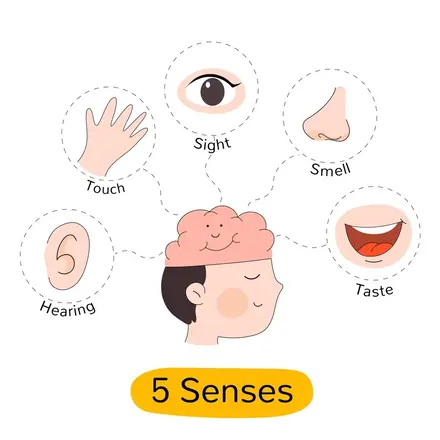
Have you ever marveled at how you experience the vibrant sights, melodious sounds, tantalizing smells, delightful tastes, and soothing touches that make life so rich? It's all thanks to the intricate workings of your five sense organs. The eyes, ears, nose, tongue, and skin are the gateways through which you perceive and interact with the world around you. These remarkable human sense organs not only enrich your daily experiences but also play a vital role in keeping you safe and healthy. So how exactly do the five sense organs work together to help you navigate and enjoy your environment? Let's take a closer look. What are the sense organs? The human body has five sense organs that allow us to perceive and interact with the environment. These include the eyes (sight), ears (hearing), nose (smell), tongue (taste), and skin (touch). Each of these organs plays a crucial role in gathering information and sending signals to the brain for processing. Sense organ functions vary based on the stimuli they detect. The eyes capture light and colour, helping us see. The ears process sound waves and maintain balance. The nose detects different odours, while the tongue identifies sweet, sour, salty, bitter, and umami tastes. The functions of the skin are to respond to temperature, pressure, and pain, allowing us to feel sensations. Together, these five types of sense organs help us navigate daily life and stay aware of our surroundings. Functions of the Five Sense Organs Now that we know what the main human sense organs are, let's explore in more detail how each one works to provide a specific sense. Sense organ functions are essential for perceiving the world, as each organ detects different types of stimuli and transmits signals to the brain. While they have distinct roles, the five sense organs often work together, sending integrated information to the brain to create a unified perceptual experience. This coordination allows us to respond effectively to our surroundings and enhances our ability to interact with the environment. Eyes: Sense of Sight Your eyes are your windows to the visual world. But have you ever wondered how they transform light into the colourful, detailed images you see? It all starts when light enters the eye through the clear, dome-shaped cornea. The light then passes through your pupil, the opening in the centre of the coloured iris, and is focused by the lens onto the retina at the back of the eye. The retina is lined with millions of light-sensitive cells called rods and cones. Rods enable you to see in dim light, while cones allow you to perceive colors and fine details. These photoreceptors convert the light into electrical signals, which then travel along the optic nerve to the visual cortex in the brain, where the image is processed and interpreted. Incredibly, the sense organs of sight—the eyes—are capable of distinguishing about 10 million different colours! However, many eye conditions can impair this vital sense, such as nearsightedness, farsightedness, and astigmatism. Regular eye check-ups can help detect and manage such issues early, preserving your precious gift of sight. Ears: Sense of Hearing From the chirping of birds to the laughter of loved ones, your ears open up the world of sound. But the process of hearing is a fascinating sequence of events. It begins when sound waves enter the ear canal and cause the eardrum to vibrate. These vibrations are then amplified by the three smallest bones in the body: the malleus (hammer), incus (anvil), and stapes (stirrup) in the middle ear. The stapes transfer the amplified vibrations to the cochlea, a fluid-filled, snail-shaped structure in the inner ear. Rippling through the cochlear fluid, the vibrations bend tiny hair cells, triggering electrical signals that travel via the auditory nerve to the brain. The brain then processes these signals, allowing you to perceive sounds and their direction, volume, and pitch. Remarkably, the sense organs responsible for hearing—the ears—can discern about 400,000 distinct sounds! However, exposure to loud noises, infections, aging, and certain medications can damage the delicate hair cells in the cochlea, leading to hearing loss. Nose: Sense of Smell From the aroma of freshly brewed coffee to the fragrance of blooming roses, your nose lets you experience the world of scents. Olfaction, or the sense of smell, begins when odour molecules enter the nostrils and dissolve in the mucus lining the roof of each nasal cavity. Embedded in this lining are millions of olfactory receptor cells, each with hair-like cilia that bind to specific odor molecules. This binding triggers receptor cells to send electrical impulses via the olfactory nerve to the olfactory bulbs, which relay the signals to the brain's olfactory cortex. The brain then identifies the smell by matching the pattern of activated receptors with previously learned odor patterns. Interestingly, your sense of smell is closely linked with your memory and emotions, which is why certain scents can vividly evoke past experiences and feelings. Did you know that the nose—one of the five sense organs—can detect over 1 trillion distinct smells? However, smoking, aging, head injuries, and certain illnesses like Parkinson's disease and Alzheimer's can impair this sensory superpower. Tongue: Sense of Taste The tantalising world of taste is brought to you by the thousands of taste buds dotting your tongue's surface. Each bud consists of 50–100 specialised gustatory receptor cells that detect the five basic tastes: sweet, salty, sour, bitter, and umami (savory). When food chemicals dissolve in saliva and interact with these receptors, electrical signals are generated and transmitted via facial nerves to the brain's gustatory cortex for identification. However, what you perceive as the "flavour" of food is actually a combination of tastes detected by your tongue and aromas detected by your nose. This is why your sense of taste may seem diminished when you have a stuffy nose. In fact, the tongue is much more limited in its taste perception compared to the nose's smell capacity, distinguishing only about 5–6 taste categories. Some factors that can affect your sense of taste include smoking, nutritional deficiencies, oral infections, and certain medications. If you experience persistent changes in how food tastes, consult your doctor to rule out any underlying issues. Skin: Sense of Touch As the body's largest sensory organ, your skin allows you to experience a wide range of tactile sensations, from the warmth of the sun to the softness of a fluffy blanket. Sense organ functions are vital for detecting and interpreting different stimuli, and the skin achieves this through a complex network of nerve endings and specialised receptors that detect touch, pressure, vibration, temperature, and pain. For example, Meissner's corpuscles respond to light touch, Pacinian corpuscles detect pressure and vibration, Merkel's discs perceive sustained pressure, and Ruffini endings sense skin stretching. When these receptors are stimulated, they send electrical signals through sensory nerves to the brain's somatosensory cortex, which processes the information and generates the appropriate touch perception. While incredibly sensitive, the skin's sensory acuity varies across different body parts. For instance, fingertips and lips are much more touch-sensitive than the back or legs, which is why using fingers to explore objects provides the most detailed tactile information. Other Sense Organs While we often focus on the traditional five senses, there are additional sensory systems that play crucial roles in our perception and interaction with the environment. Two of these lesser-known sense organs are the vestibular system, responsible for balance and coordination, and the proprioception system, which provides body awareness. The vestibular system, located in the inner ear, helps us maintain stability and spatial orientation. Proprioception, often called the "sixth sense," allows us to perceive the position and movement of our body parts without looking. These sense organs' functions are essential for smooth movement, posture control, and overall physical coordination. Vestibular System (Balance and Coordination) The vestibular system, located in the inner ear, is essential for maintaining balance and spatial orientation. It consists of the otolith organs (utricle and saccule) and the semicircular canals, which detect changes in head position and movement. When you tilt your head, spin around, or suddenly stop, the vestibular system helps you maintain equilibrium and prevents dizziness. It works closely with the visual and proprioceptive systems to ensure smooth movement and coordination. An interesting fact: The vestibular system is so sensitive that it can detect head movements as small as 0.004 degrees! This incredible precision allows us to stay steady while walking, running, or even standing still in a moving environment. Proprioception System (Body Awareness) Have you ever wondered how you can touch your nose with your eyes closed or walk without looking at your feet? This is thanks to the proprioception system, which allows us to sense the position and movement of our body parts without visual input. Proprioceptors, located in muscles, tendons, and joints, constantly send information to the brain about the relative positioning of our limbs. Proprioception enables us to perform complex tasks, like playing musical instruments or typing on a keyboard, with incredible precision and coordination. It's like having a sixth sense that keeps track of our body's movements and adjusts muscle tension and joint positions accordingly. How to Keep Sense Organs Healthy? Maintaining the health of our sense organs is crucial for ensuring optimal sensory function throughout our lives. Our eyes, ears, nose, skin, and tongue help us perceive the world, and taking proper care of them ensures their longevity and efficiency. Here are essential tips for keeping each of the five sense organs healthy: Eye Care Tips The eyes are one of the most vital sense organs, allowing us to see and interpret the world around us. Keeping them healthy ensures good vision and prevents eye-related diseases. Schedule Regular Eye Exams: Visit an eye specialist at least once a year to detect and address any vision problems early on. Early diagnosis of conditions like cataracts, glaucoma, and macular degeneration can prevent severe damage. Wear Protective Eyewear: Use safety goggles or protective glasses when working with chemicals, sharp objects, or engaging in sports to prevent injuries. Maintain a Nutritious Diet: Consume foods rich in vitamins A and C, as well as omega-3 fatty acids, to support eye health. Carrots, spinach, citrus fruits, and fish are excellent choices. Reduce Screen Time: Prolonged exposure to screens can cause eye strain and fatigue. Follow the 20-20-20 rule: Every 20 minutes, look at something 20 feet away for 20 seconds. Ear Care Tips Our ears help us hear and maintain balance. Taking care of them can prevent hearing loss and other ear-related problems. Protect Against Loud Noises: Avoid prolonged exposure to loud music or noise. Use earplugs or noise-canceling headphones in noisy environments. Avoid Inserting Objects in the Ear: Refrain from using cotton swabs or any sharp objects inside your ears, as they can damage the eardrum or push wax deeper. Get Regular Hearing Tests: Hearing tests can help detect early signs of hearing loss. Consult an audiologist if you experience ringing in the ears (tinnitus) or difficulty hearing. Limit Use of Earphones: Using earphones at high volumes can damage your hearing. Keep the volume at 60% or lower and take breaks. Nose and Smell Care Tips The nose plays a crucial role in breathing and detecting scents. Keeping it healthy ensures a strong sense of smell and clear nasal passages. Keep Nasal Passages Moisturised: Use saline sprays or a humidifier, especially in dry environments, to prevent nasal dryness. Avoid Strong Chemicals and Pollutants: Exposure to harsh chemicals, smoke, or strong odors can damage olfactory receptors. Wear a mask if necessary. Quit Smoking: Smoking can dull your sense of smell and lead to respiratory problems. Practice Good Hygiene: Regularly clean your hands and avoid touching your nose to prevent infections like colds and sinusitis. Skin Care Tips The skin is the largest sense organ and is responsible for touch, temperature detection, and protection from external elements. Use Sunscreen: Apply sunscreen with an adequate SPF to protect against harmful UV rays that can cause premature aging and skin cancer. Keep Skin Moisturised: Dry skin can become irritated and lose sensitivity. Use a good-quality moisturiser to keep your skin healthy and hydrated. Be Gentle When Cleansing: Avoid harsh soaps and scrubbing too hard, as this can damage the skin barrier and reduce tactile sensitivity. Stay Hydrated: Drink plenty of water to maintain skin elasticity and overall health. Eat a Balanced Diet: Consume foods rich in vitamins E and C to promote healthy skin. Tongue and Taste Care Tips The tongue helps us taste and digest food. Keeping it clean and healthy ensures an optimal sense of taste and prevents infections. Maintain Good Oral Hygiene: Brush your teeth twice daily, floss regularly, and clean your tongue to prevent bacterial buildup. Stay Hydrated: A dry mouth can reduce your ability to taste. Drinking enough water keeps the tongue moist and functioning properly. Consume a Balanced Diet: Zinc, iron, and vitamin B12 are essential for maintaining taste bud health. Include nuts, lean meat, and dairy in your diet. Avoid Excessive Sugar and Spicy Foods: Too much sugar can cause oral infections, while very spicy foods can irritate taste buds. Avoid Smoking and Alcohol: These substances can dull your sense of taste over time. Frequently Asked Questions What is the function of sense organs? Sense organs are specialised structures that allow us to perceive and interact with the world around us. They gather information about our environment, such as sights, sounds, smells, tastes, and tactile sensations, and transmit this information to the brain for interpretation. Sense organs functions are essential for our survival, safety, and enjoyment of life. Which is the largest sense organ? The skin is the largest sense organ in the human body. It is responsible for the sense of touch, allowing us to perceive temperature, pressure, pain, and texture. The skin covers an impressive surface area of approximately 2 square meters in adults, making it the most extensive of the five sense organs. Can a person have no sense of touch? While extremely rare, there are conditions that can impair or eliminate a person's sense of touch. For example, individuals with certain neurological disorders or nerve damage may experience reduced or absent tactile sensation. However, a complete lack of touch sensation is highly uncommon and would significantly impact an individual's ability to interact with their environment safely. What are olfactory organs? Olfactory organs refer to the structures in the nose responsible for the sense of smell. The primary olfactory organ is the olfactory epithelium, which contains millions of olfactory receptors. These receptors detect odor molecules in the air and send signals to the brain, allowing us to perceive and distinguish between different scents. Which part of the human ear maintains body balance? The vestibular apparatus, located in the inner ear, is responsible for maintaining body balance and spatial orientation. It consists of the otolith organs (utricle and saccule) and the semicircular canals, which detect changes in head position and movement. The vestibular system works in conjunction with other sense organs, such as the eyes and proprioceptors, to keep us balanced and coordinated. Conclusion The five sense organs - eyes, ears, nose, tongue, and skin - along with the vestibular and proprioception systems, form an incredible network that allows us to perceive, interact with, and enjoy the world around us. By understanding how these sense organs work together and taking steps to maintain their health, we can ensure a more complete and fulfilling sensory experience throughout our lives. At Metropolis Healthcare, we understand the importance of maintaining healthy sense organs. Our team of expert pathologists and technicians provide accurate and reliable diagnostic services, including tests related to sensory health. With our convenient at-home sample collection and user-friendly online reporting system, prioritising your well-being has never been easier.














 WhatsApp