


Latest Blogs
दालचिनीचे फायदे: विज्ञानाने समर्थित 12 प्रभावी आरोग्यदायी फायदे
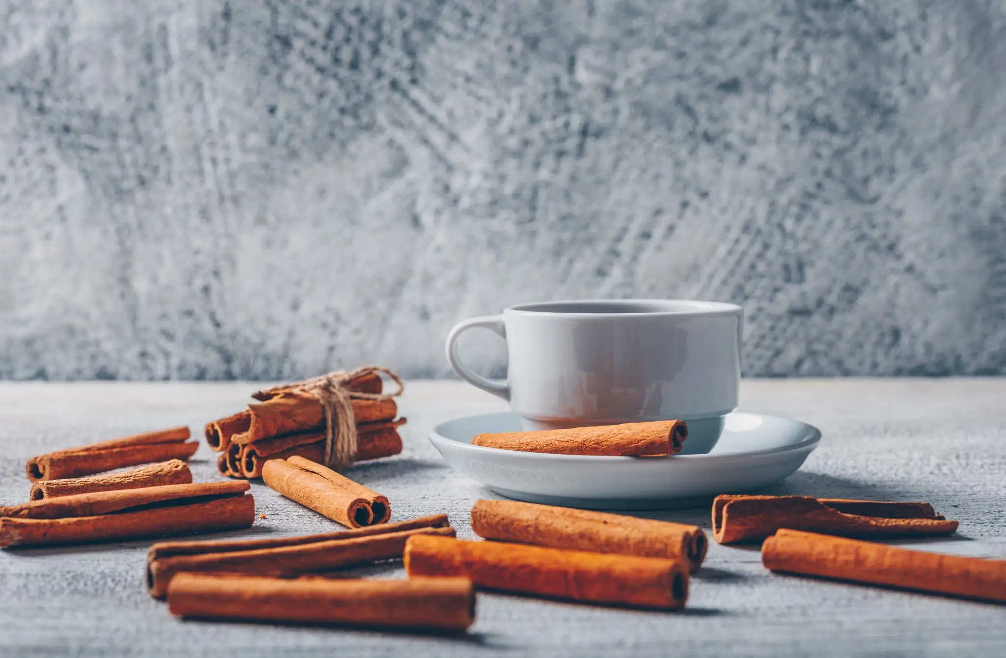
दालचिनी म्हणजे काय? दालचिनी हा एक सुगंधी मसाला आहे, जो सिनॅमोमम कुटुंबातील सदाहरित झाडांच्या आतील सालीपासून तयार केला जातो. हा आवडता मसाला दालचिनीच्या झाडापासून मिळतो, जे मुख्यतः आशिया आणि मध्य पूर्वेतील उष्णकटिबंधीय प्रदेशांमध्ये वाढते. ही साल काळजीपूर्वक काढली जाते, वाळवली जाते आणि नैसर्गिकरित्या आपल्याला परिचित असलेल्या वैशिष्ट्यपूर्ण गुंडाळीदार काड्यांच्या आकारात वळते. जगभरात दालचिनीचे दोन मुख्य प्रकार उपलब्ध आहेत. सिलोन दालचिनी, जी अनेकदा "खरी दालचिनी" म्हणून ओळखली जाते, तिचा उगम श्रीलंकेत झाला असून तिची चव सौम्य आणि गोडसर असते. कॅशिया दालचिनी — जी भारतीय घरांमध्ये दालचिनी म्हणून मोठ्या प्रमाणावर ओळखली जाते — हिची चव अधिक ठसठशीत आणि तीव्र असते आणि स्वयंपाक व पारंपरिक औषधोपचारांमध्ये सर्वाधिक वापरला जाणारा हा प्रकार आहे. दालचिनीचे दोन्ही प्रकार आरोग्यदायी फायदे देतात, मात्र सिलोन दालचिनीमध्ये कूमरिनचे प्रमाण कमी असल्याने ती नियमित सेवनासाठी अधिक सुरक्षित मानली जाते. दालचिनीच्या फायद्यांसाठी जबाबदार जैवसक्रिय घटकांमध्ये सिनामाल्डिहाइड, सिनॅमिक आम्ल आणि विविध पॉलीफेनॉल अँटिऑक्सिडंट्स यांचा समावेश होतो, जे या मसाल्याला उपचारात्मक गुण देतात. दालचिनीचे पोषणमूल्य यूएसडीएनुसार, खालील तक्त्यात 100 g दळलेल्या दालचिनीतील पोषणघटकांचे प्रमाण दाखवले आहे: पोषक घटक प्रमाण (per 100g) ऊर्जा 247 kcal प्रथिने 3.99 g एकूण चरबी 1.24 g कार्बोहायड्रेट 80.6 g एकूण तंतू 53.1 g एकूण साखर 2.17 g कॅल्शियम 1000 mg लोह 8.32 mg मॅग्नेशियम 60 mg फॉस्फरस 64 mg पोटॅशियम 431 mg सोडियम 10 mg झिंक 1.83 mg मॅंगनीज 17.5 mg व्हिटॅमिन C 3.8 mg व्हिटॅमिन B6 0.158 mg व्हिटॅमिन A 295 IU व्हिटॅमिन K 31.2 µg दालचिनीचे 12 प्रभावी आरोग्यदायी फायदे 1. संसर्गांपासून संरक्षण करते दालचिनीमध्ये उल्लेखनीय जंतुरोधक गुणधर्म आढळतात, ज्यामुळे ती विविध रोगकारकांपासून नैसर्गिक संरक्षण देते. दालचिनीतील सक्रिय घटक हानिकारक जीवाणू, बुरशी (कँडिडा प्रजातींसह) आणि काही विषाणूंची वाढ रोखू शकतात. हा संरक्षक परिणाम तोंडाच्या आरोग्यापर्यंतही पोहोचतो, जिथे दालचिनी दातांमध्ये किड आणि हिरड्यांचे विकार निर्माण करणाऱ्या जीवाणूंशी लढण्यास मदत करू शकते. सकाळच्या चहात दालचिनी पावडर घालणे किंवा आहारात दालचिनीचे पाणी समाविष्ट करणे यामुळे तोंडातील संसर्गांपासून अतिरिक्त संरक्षण मिळू शकते आणि श्वासही नैसर्गिकरीत्या ताजा राहू शकतो. 2. रक्तातील साखर नियंत्रित ठेवते दालचिनीच्या सर्वाधिक अभ्यासलेल्या फायद्यांपैकी एक म्हणजे रक्तातील साखरेचे नियमन. संशोधनात दिसून आले आहे की दालचिनीचे नियमित सेवन केल्याने उपाशीपोटी रक्तातील ग्लुकोजची पातळी लक्षणीयरीत्या कमी होऊ शकते आणि इन्सुलिन संवेदनशीलता सुधारू शकते, ज्याचा विशेष फायदा टाइप 2 मधुमेह किंवा मेटाबॉलिक सिंड्रोम असलेल्या व्यक्तींना होऊ शकतो. अभ्यासानुसार दररोज साधारण अर्धा चमचा दालचिनी पावडर घेतल्यास रक्तातील ग्लुकोज नियंत्रणात अर्थपूर्ण सुधारणा दिसू शकते. हे जठर रिकामे होण्याची गती कमी करून आणि पेशींमध्ये ग्लुकोजचे शोषण वाढवून कार्य करते, ज्यामुळे जेवणानंतर रक्तातील साखरेत होणारी धोकादायक वाढ टाळण्यास मदत होते. 3. कोलेस्टेरॉल कमी करते संशोधनानुसार दालचिनी एकूण कोलेस्टेरॉल, हानिकारक LDL कोलेस्टेरॉल आणि ट्रायग्लिसराइड्स कमी करण्यास मदत करू शकते, तसेच उपयुक्त HDL कोलेस्टेरॉल वाढवण्यासही मदत करू शकते. हे परिणाम दालचिनी यकृतातील चरबीच्या चयापचयाला मदत करते आणि आहारातून कोलेस्टेरॉलचे शोषण कमी करते म्हणून दिसून येतात. कोलेस्टेरॉलची पातळी वाढलेली असलेल्या व्यक्तींनी निरोगी आहारासोबत दैनंदिन दिनचर्येत दालचिनीचा समावेश केल्यास चांगला फायदा होऊ शकतो. कोलेस्टेरॉल कमी करण्यासाठी दालचिनीचे फायदे विशेषतः तेव्हा अधिक स्पष्ट दिसतात, जेव्हा तिचे सेवन अनेक महिन्यांपर्यंत सातत्याने केले जाते. त्यामुळे हृदयविकारांपासून बचावासाठी ही दीर्घकालीन चांगली पद्धत ठरू शकते. 4. हृदयाच्या आरोग्यास मदत करते दालचिनी रक्तदाब कमी करण्यास, रक्तवाहिन्यांतील सूज कमी करण्यास आणि हृदयरोगाशी संबंधित ऑक्सिडेटिव्ह ताणापासून संरक्षण देण्यास मदत करू शकते. दालचिनी पावडरमधील शक्तिशाली अँटिऑक्सिडंट्स धमन्यांना इजा पोहोचवणाऱ्या फ्री रॅडिकल्सना निष्क्रिय करतात, तर तिचे दाहरोधक घटक रक्तवाहिन्यांचे आरोग्य टिकवून ठेवण्यास मदत करतात. याशिवाय, दालचिनीच्या पाण्याचे फायदे म्हणजे रक्ताभिसरणाला मदत होणे आणि कदाचित रक्ताच्या गाठी तयार होण्याचा धोका कमी होणे. अभ्यासानुसार नियमितपणे दालचिनीचे सेवन करणाऱ्या लोकांमध्ये हृदयरोग होण्याचा धोका कमी असू शकतो, विशेषतः जेव्हा ती इतर हृदयासाठी हितकारक जीवनशैलीच्या सवयींसोबत घेतली जाते. 5. सूज कमी करते दीर्घकालीन सूज अनेक आरोग्यसमस्यांच्या मुळाशी असते आणि दालचिनीतील प्रभावी दाहरोधक घटक उपचारात्मक दृष्टीने महत्त्वाची क्षमता दर्शवतात. या मसाल्यात सिनामाल्डिहाइड आणि इतर जैवसक्रिय घटक असतात, जे शरीरभर होणाऱ्या दाहप्रतिक्रियांना नियंत्रित करण्यात मदत करू शकतात. हा दाहरोधक परिणाम संधिवात यांसारख्या स्थितींमध्ये विशेषतः उपयुक्त ठरू शकतो, कारण सूज कमी झाल्यास जीवनमान आणि सांध्यांची कार्यक्षमता लक्षणीयरीत्या सुधारू शकते. 6. आतड्यातील उपयुक्त जीवाणूंचा समतोल राखते संशोधनानुसार दालचिनीमध्ये प्रीबायोटिकसारखे घटक असतात, जे पचनसंस्थेच्या आरोग्यास मदत करतात. हे उपयुक्त आतडीतील जीवाणूंना वाढण्यास प्रोत्साहन देतात आणि हानिकारक सूक्ष्मजीवांना मर्यादित ठेवतात. तिचा सक्रिय घटक सिनामाल्डिहाइड आतड्यातील मायक्रोबायोममध्ये सकारात्मक बदल घडवून आणण्यास, शॉर्ट-चेन-फॅटी-अॅसिड निर्माण करणारे जीवाणू वाढवण्यास आणि आतड्यांच्या संरक्षक आवरणाला बळकटी देण्यास मदत करू शकतो. या सर्व संयुक्त परिणामांमुळे पचनक्रिया सुधारू शकते आणि जठरांत्रातील अस्वस्थता कमी होऊ शकते. 7. अँटिऑक्सिडंट्सने समृद्ध आहे दालचिनीमध्ये पॉलीफेनॉल्स आणि फ्लॅव्होनॉइड्ससारखे शक्तिशाली घटक असतात, जे तुमच्या पेशींना ऑक्सिडेटिव्ह इजापासून संरक्षण देतात. हे अँटिऑक्सिडंट्स वृद्धत्व आणि दीर्घकालीन आजारांना कारणीभूत ठरणाऱ्या फ्री रॅडिकल्सना निष्क्रिय करतात. दालचिनी पावडरमधील उच्च अँटिऑक्सिडंट प्रमाण हे विशेषतः कर्करोग, हृदयरोग आणि न्यूरोडिजेनेरेटिव्ह स्थितींना कारणीभूत ठरू शकणाऱ्या पेशींच्या नुकसानीपासून संरक्षण देण्यासाठी महत्त्वाचे ठरते. तिचे नियमित सेवन शरीराच्या नैसर्गिक संरक्षणव्यवस्थांना मदत करते. 8. मज्जासंस्थेचे संरक्षण करणारे परिणाम देते संशोधन दर्शवते की दालचिनीमध्ये अँटिऑक्सिडंट, दाहरोधक आणि पेशीमृत्यू-रोधक गुणधर्म असल्यामुळे ती मज्जासंस्थेचे संरक्षण करणारे फायदे देते. सिनामाल्डिहाइड आणि सोडियम बेंझोएट यांसारखे घटक मज्जापेशींना ऑक्सिडेटिव्ह ताणापासून वाचवण्यास, न्यूरोट्रान्समिटर्सचे नियमन करण्यास आणि पेशीमृत्यू रोखण्यास मदत करतात. प्रथिनांचे एकत्रीकरण कमी करून आणि मज्जापेशींची स्थिरता टिकवून दालचिनी मेंदूच्या आरोग्यासाठी नैसर्गिक सहाय्यक ठरू शकते, विशेषतः पार्किन्सन रोग, अल्झायमर रोग आणि मल्टिपल स्क्लेरोसिस यांसारख्या स्थितींमध्ये. 9. कर्करोगाचा धोका कमी करण्यात मदत करू शकते प्रयोगशाळेतील अभ्यासांमधून दिसून आले आहे की दालचिनीतील घटक, विशेषतः सिनामाल्डिहाइड, ट्यूमरची वाढ रोखणे आणि कर्करोगी पेशींचा मृत्यू वाढवणे अशा प्रकारे कर्करोगविरोधी गुणधर्म दर्शवतात. प्रारंभिक संशोधनात हे परिणाम विविध प्रकारच्या कर्करोगांमध्ये आढळले आहेत. दालचिनीचे अँटिऑक्सिडंट गुणधर्म कर्करोगाच्या विकासाला कारणीभूत ठरू शकणाऱ्या DNA नुकसानीपासून संरक्षण देतात, तर तिचे दाहरोधक परिणाम कर्करोगाच्या धोक्याशी संबंधित दीर्घकालीन सूज कमी करण्यास मदत करू शकतात. तथापि, हे निष्कर्ष आशादायक असले तरी, कर्करोग प्रतिबंधामध्ये दालचिनीची भूमिका पूर्णपणे समजून घेण्यासाठी मानवांवरील अधिक अभ्यासांची आवश्यकता आहे. 10. रोगप्रतिकारक शक्तीला मदत करते दालचिनीतील जंतुरोधक आणि दाहरोधक गुणधर्म रोगप्रतिकारक शक्ती अधिक सक्षम ठेवण्यास मदत करू शकतात. दालचिनीतील व्हिटॅमिन C आणि इतर पोषक घटक पांढऱ्या रक्तपेशींच्या कार्याला मदत करतात, तर तिचे जंतुरोधक घटक रोगकारक सूक्ष्मजीवांची वाढ रोखण्यास हातभार लावतात. नियमित सेवनामुळे तुमचे शरीर संसर्गांपासून अधिक चांगल्या प्रकारे बचाव करू शकते आणि योग्य रोगप्रतिकारक प्रतिसाद टिकवून ठेवू शकते. 11. वजन नियंत्रणात मदत करते दालचिनी अनेक मार्गांनी निरोगी वजन नियंत्रणाला मदत करू शकते. ती रक्तातील साखरेची पातळी नियंत्रित ठेवते, ज्यामुळे भूक आणि काही खाण्याची इच्छा कमी होऊ शकते तसेच जास्त खाण्याकडे नेणारे ऊर्जा-पातळीतील अचानक घसरणे टाळता येते. याशिवाय, दालचिनी चयापचय वाढवण्यास आणि शरीराला चरबी अधिक कार्यक्षमतेने जाळण्यास मदत करू शकते. हा मसाला जठर रिकामे होण्याची गतीही कमी करतो, ज्यामुळे जेवणानंतर जास्त वेळ पोट भरल्यासारखे वाटू शकते. 12. अॅलर्जीपासून आराम मिळविण्यास मदत करते दालचिनीतील दाहरोधक आणि रोगप्रतिकारक प्रतिसाद नियंत्रित करणारे गुणधर्म अॅलर्जिक प्रतिक्रिया कमी करण्यात मदत करू शकतात. हिस्टामीनसारख्या दाहकारक घटकांचे उत्सर्जन रोखून दालचिनी ऋतुमानानुसार होणाऱ्या अॅलर्जी आणि इतर अॅलर्जिक प्रतिक्रियांपासून आराम देऊ शकते. दालचिनीचे नियमित सेवन अॅलर्जीच्या काळात शिंक, नाक बंद होणे आणि डोळ्यांना खाज येणे यांसारखी लक्षणे कमी करण्यात मदत करू शकते. दालचिनीतील अँटिऑक्सिडंट्स अॅलर्जिक सूजेशी संबंधित ऑक्सिडेटिव्ह ताण कमी करण्यासही मदत करतात. दालचिनीने पारंपरिक अॅलर्जी उपचारांची जागा घेऊ नये, मात्र सौम्य अॅलर्जिक लक्षणांचे व्यवस्थापन करण्यासाठी ती पूरक आधार देऊ शकते. दालचिनीचे संभाव्य दुष्परिणाम आणि घ्यायची काळजी दालचिनीचे स्वयंपाकातील प्रमाणात सेवन बहुतेक लोकांसाठी सामान्यतः सुरक्षित असले तरी, संभाव्य दुष्परिणाम आणि घ्यायची काळजी यांची जाणीव असणे महत्त्वाचे आहे. कॅशिया दालचिनीचे अतिसेवन तिच्यातील जास्त कूमरिनमुळे यकृताचे नुकसान करू शकते. सिलोन दालचिनीमध्ये कूमरिनचे प्रमाण खूपच कमी असते आणि ती नियमित वापरासाठी अधिक सुरक्षित असते. काही व्यक्तींना, विशेषतः इतर मसाल्यांबद्दल संवेदनशीलता असल्यास, अॅलर्जिक प्रतिक्रिया येऊ शकतात. दालचिनीचे मोठ्या प्रमाणात सेवन केल्यास, विशेषतः दालचिनीच्या गाढ तेलामुळे, तोंडात जखमा किंवा जळजळ होऊ शकते. जर तुम्ही रक्त पातळ करणारी औषधे किंवा मधुमेहाची औषधे घेत असाल, तर दालचिनीचे सेवन लक्षणीयरीत्या वाढवण्यापूर्वी डॉक्टरांचा सल्ला घ्या, कारण ती या औषधांचा परिणाम वाढवू शकते. गर्भवती किंवा स्तनपान करणाऱ्या महिलांनी दालचिनीचे सेवन स्वयंपाकातील प्रमाणापुरते मर्यादित ठेवावे आणि वैद्यकीय सल्ल्याशिवाय गाढ पूरक स्वरूपातील उत्पादने टाळावीत. निष्कर्ष वैज्ञानिक संशोधनाने समर्थित दालचिनीचे विस्तृत फायदे हा सुगंधी मसाला तुमच्या आरोग्यदायी दिनचर्येत महत्त्वाची भर ठरवतात. रक्तातील साखरेचे नियमन आणि हृदयाच्या आरोग्याला मदत करण्यापासून ते रोगप्रतिकारक शक्ती बळकट करणे आणि सूज कमी करणे यापर्यंत, दालचिनी स्वादिष्ट स्वरूपात उल्लेखनीय उपचारात्मक क्षमता देते. तुम्ही स्वयंपाकात दालचिनी पावडर वापरत असाल, दालचिनीच्या पाण्याचे फायदे घेत असाल किंवा फक्त सकाळच्या कॉफीत ती घालत असाल, तर दररोजचे छोटे प्रमाणदेखील तुमच्या आरोग्याच्या उद्दिष्टांसाठी महत्त्वाचे योगदान देऊ शकते. शक्य असल्यास नियमित सेवनासाठी सिलोन दालचिनी निवडा आणि जर तुम्हाला आधीपासून काही वैद्यकीय समस्या असतील किंवा तुम्ही औषधे घेत असाल, तर नेहमी डॉक्टरांचा सल्ला घ्या. मेट्रोपोलिस हेल्थकेअरमध्ये आम्हाला हे समजते की दैनंदिन जीवनात दालचिनीचा समावेश करण्यासारख्या नैसर्गिक आरोग्यपद्धती वापरताना तुमच्या आरोग्य निर्देशकांवर लक्ष ठेवणे किती महत्त्वाचे आहे. आमच्या 4,000 पेक्षा अधिक तपासण्यांच्या व्यापक संचात रक्तातील साखरेचे निरीक्षण, कोलेस्टेरॉल तपासणी आणि दाह निर्देशकांसाठी विशेष पॅनेल्सचा समावेश आहे, जे तुमची प्रगती समजून घेण्यास मदत करू शकतात. भारतभरातील 10,000+ टचपॉइंट्समध्ये उपलब्ध आमच्या सोयीस्कर घरपोच नमुना संकलन सेवेमुळे तुम्हाला घरबसल्या तुमच्या आरोग्यप्रवासाला अधिक योग्य बनवण्यासाठी आवश्यक निदानविषयक माहिती सहज मिळू शकते. सामान्य प्रश्न दालचिनीचे आरोग्यदायी फायदे कोणते आहेत? दालचिनीचे विज्ञानाधारित अनेक फायदे आहेत, ज्यात रक्तातील साखरेचे नियमन, कोलेस्टेरॉल कमी करणे, दाहरोधक परिणाम, अँटिऑक्सिडंट संरक्षण, रोगप्रतिकारक शक्तीला मदत आणि नियमितपणे मर्यादित प्रमाणात सेवन केल्यास हृदयविषयक संभाव्य फायदे यांचा समावेश होतो. दालचिनी वजन कमी करण्यात मदत करू शकते का? होय, दालचिनी रक्तातील साखरेची पातळी स्थिर ठेवणे, भूक आणि खाण्याची इच्छा कमी करणे, जठर रिकामे होण्याची गती कमी करणे आणि निरोगी आहार व व्यायामासोबत घेतल्यास कदाचित चयापचय वाढवणे अशा मार्गांनी वजन नियंत्रणाला मदत करू शकते. रक्तातील साखरेवर नियंत्रण ठेवण्यासाठी दालचिनी उपयुक्त आहे का? संशोधनाने पुष्टी केली आहे की दालचिनी इन्सुलिन संवेदनशीलता सुधारू शकते आणि उपाशीपोटी रक्तातील ग्लुकोजची पातळी कमी करू शकते. दररोज अर्धा ते दोन चमचे इतक्या प्रमाणात सातत्याने सेवन केल्यास याचा विशेष फायदा टाइप 2 मधुमेह असलेल्या लोकांना होऊ शकतो. दालचिनी पचनासाठी मदत करते का? दालचिनी आतड्यातील उपयुक्त जीवाणूंचा समतोल राखण्यास, हानिकारक सूक्ष्मजीव कमी करण्यास आणि तिच्या जंतुरोधक व दाहरोधक गुणधर्मांमुळे वायू आणि पोटफुगी कमी करण्यास मदत करून पचनसंस्थेच्या आरोग्यास लाभदायी ठरू शकते. दालचिनीचे सेवन केल्याने काही दुष्परिणाम होऊ शकतात का? कॅशिया दालचिनीचे अतिसेवन केल्याने यकृताला विषारी परिणाम होऊ शकतो संवेदनशील व्यक्तींमध्ये अॅलर्जिक प्रतिक्रिया होऊ शकतात गाढ स्वरूपातील दालचिनीमुळे तोंडात जळजळ होऊ शकते मधुमेहाच्या औषधांसोबत घेतल्यास रक्तातील साखर धोकादायकरीत्या कमी होऊ शकते रक्त पातळ करणाऱ्या औषधांसोबत संभाव्य औषध-परस्परसंवाद होऊ शकतो मी माझ्या आहारात दालचिनीचा समावेश कसा करू शकतो? ओट्स, दही किंवा ताज्या फळांवर दालचिनी पावडर शिंपडा अधिक चवीसाठी स्मूदी, कॉफी किंवा चहात घाला बेकिंगच्या पाककृतींमध्ये आणि गोड पदार्थांमध्ये वापरा कोमट पाण्यात पावडर मिसळून दालचिनीचे पाणी तयार करा करी आणि भाताच्या पदार्थांसारख्या चविष्ट पदार्थांमध्ये वापरा विविध पाककृतींसाठी घरच्या घरी मसाल्याचे मिश्रण तयार करा सिलोन दालचिनी कॅशिया दालचिनीपेक्षा चांगली आहे का? सिलोन दालचिनीमध्ये कूमरिनचे प्रमाण लक्षणीयरीत्या कमी असल्यामुळे ती नियमित सेवनासाठी सामान्यतः अधिक सुरक्षित मानली जाते. कूमरिन हा एक असा घटक आहे, जो मोठ्या प्रमाणात घेतल्यास यकृताचे नुकसान करू शकतो. मात्र, योग्य प्रकारे वापरल्यास दोन्ही प्रकारच्या दालचिनी आरोग्यदायी फायदे देतात.
डीएचटी ब्लॉकर अन्नपदार्थ: केसांची वाढ समर्थित करण्यासाठी आणि केस गळणे टाळण्यासाठी 12 नैसर्गिक मार्ग
डीएचटी म्हणजे काय आणि त्यामुळे केस का गळतात? डायहायड्रोटेस्टोस्टेरॉन (डीएचटी) हा एक प्रभावी हार्मोन आहे, जो टेस्टोस्टेरॉनपासून 5-अल्फा-रिडक्टेस (5-alpha-reductase) नावाच्या एन्झाइमच्या क्रियेने तयार होतो. ज्यांना आनुवंशिकदृष्ट्या प्रवृत्ती असते अशा व्यक्तींमध्ये, डीएचटी केसांच्या फॉलिकल्सवरील रिसेप्टर्सना चिकटतो, त्यांच्या नैसर्गिक वाढीच्या चक्रात अडथळा आणतो आणि केसांच्या आरोग्यात लक्षणीय बदल घडवतो. जेव्हा डीएचटी केसांच्या फॉलिकल्सना चिकटतो, तेव्हा तो वाढीचा टप्पा कमी करतो आणि विश्रांती व गळतीचे टप्पे वाढवतो. या प्रक्रियेमुळे केस हळूहळू अधिक पातळ, कमकुवत होतात आणि पुन्हा वाढण्यापेक्षा जलद गळू लागतात. कालांतराने, डीएचटी फॉलिक्युलर मिनिएचरायझेशन नावाच्या प्रक्रियेद्वारे फॉलिकल्स लहान करतो, ज्यामुळे अँड्रोजेनेटिक अलोपेशियामध्ये दिसणारा केस पातळ होण्याचा ठरावीक नमुना तयार होतो. ही प्रक्रिया समजल्यावर, आहारातील उपायांद्वारे डीएचटीवर लक्ष केंद्रित करणे केसांच्या आरोग्यास नैसर्गिकरीत्या मदत करण्यासाठी प्रभावी धोरण का ठरू शकते, हे स्पष्ट होते. डीएचटी ब्लॉकर अन्नपदार्थ कसे कार्य करतात: 5-अल्फा-रिडक्टेस रोखण्याची क्रिया सर्वोत्तम डीएचटी ब्लॉकर अन्नपदार्थांमध्ये विशिष्ट पोषकद्रव्ये आणि जैवसक्रिय संयुगे असतात, जी टेस्टोस्टेरॉनचे डीएचटीमध्ये रूपांतर करणाऱ्या मुख्य 5-अल्फा-रिडक्टेस एन्झाइमच्या क्रियेला रोखू शकतात. ही रूपांतरण प्रक्रिया कमी करून, हे अन्नपदार्थ केसांच्या फॉलिकल्सच्या पातळीवर डीएचटीचे प्रमाण कमी करण्यास मदत करतात, ज्यामुळे फॉलिकल्स लहान होण्याची प्रक्रिया आणि केस गळणे कमी होण्यास मदत होऊ शकते. डीएचटी ब्लॉकर अन्नपदार्थांमध्ये आढळणारी काही नैसर्गिक संयुगे अँड्रोजन रिसेप्टरची क्रिया नियंत्रित करू शकतात किंवा निरोगी केसवाढीस मदत करणाऱ्या पेशी-संकेत मार्गांवर प्रभाव टाकू शकतात. या क्रिया एकत्रितपणे काम करून मजबूत आणि तगडे केसांचे फॉलिकल्स टिकवून ठेवण्यासाठी अधिक अनुकूल वातावरण तयार करतात. कोणतीही डीएचटी ब्लॉकर अन्नपद्धती किती परिणामकारक ठरेल हे नियमित सेवनावर आणि एका घटकावर अवलंबून न राहता अनेक उपयुक्त पोषकद्रव्ये एकत्र घेण्यावर अवलंबून असते. जगभरात डीएचटी आणि केस गळतीचा धोका वाढवणारे घटक पुराव्यावर आधारित क्लिनिकल संदर्भग्रंथ एंडोटेक्स्टमधील एका अभ्यासानुसार, 50 वर्षांवरील सुमारे 50% लोकांमध्ये केस गळण्याची समस्या दिसते आणि अनेक प्रकरणे डायहायड्रोटेस्टोस्टेरॉन (डीएचटी) च्या वाढलेल्या पातळीशी संबंधित असतात. पुरुष आणि महिलांमध्ये डीएचटीची पातळी वाढवणारे आणि केस गळतीचा धोका वाढवणारे घटक पुढीलप्रमाणे आहेत: आनुवंशिक प्रवृत्ती: अँड्रोजेनेटिक अलोपेशियाचा कौटुंबिक इतिहास धोका लक्षणीयरीत्या वाढवतो वाढते वय: वयानुसार डीएचटीप्रती संवेदनशीलता सामान्यतः वाढते पुरुष जैविक लिंग: पुरुषांमध्ये डीएचटीमुळे होणारे केसगळणे अधिक प्रमाणात दिसते हार्मोनल असंतुलन: टेस्टोस्टेरॉन किंवा डीएचटीची वाढलेली पातळी फॉलिकल्सचे नुकसान वेगाने वाढवते 5-अल्फा-रिडक्टेसची जास्त क्रिया: एन्झाइमची वाढलेली पातळी डीएचटीचे रूपांतर वाढवते पर्यावरणीय घटक: अयोग्य आहार, दीर्घकालीन ताण आणि निष्क्रिय जीवनशैली वैद्यकीय स्थिती: महिलांमध्ये पॉलीसिस्टिक ओव्हरी सिंड्रोम, पुरुषांमध्ये प्रोस्टेटच्या समस्या लठ्ठपणा आणि मेटाबॉलिक सिंड्रोम: या स्थितींमुळे हार्मोनल संतुलन बदलते अॅनाबॉलिक स्टेरॉइड्सचा वापर: कृत्रिम हार्मोन्स डीएचटीचे उत्पादन मोठ्या प्रमाणात वाढवतात पोषण आणि आहार केसांच्या आरोग्यावर आणि डीएचटीच्या पातळीवर कसा प्रभाव टाकतात आहाराचा तुमच्या शरीरातील हार्मोनल वातावरणावर, त्यात डीएचटी निर्मितीच्या पातळीवर, थेट प्रभाव पडतो. झिंक, व्हिटॅमिन B6 आणि विशिष्ट वनस्पतीजन्य पॉलीफेनॉल्स यांसारखी पोषकद्रव्ये 5-अल्फा-रिडक्टेसची क्रिया प्रभावीपणे नियंत्रित करू शकतात, ज्यामुळे शरीरभर डीएचटीची निर्मिती नैसर्गिकरीत्या कमी होऊ शकते. अँटिऑक्सिडंट्स, दाहरोधक संयुगे आणि आवश्यक फॅटी अॅसिड्सने समृद्ध संतुलित आहार ऑक्सिडेटिव्ह ताण आणि सूज यांपासून संरक्षण देऊन केसांच्या फॉलिकल्सच्या एकूण आरोग्यास मदत करतो; हे दोन्ही केसगळणे वाढवू शकतात. दीर्घकाळ पोषकद्रव्यांची कमतरता, अतिप्रक्रियायुक्त अन्नपदार्थांचे जास्त सेवन किंवा संतृप्त चरबीचे अधिक प्रमाण हार्मोनल असंतुलन वाढवू शकते, ज्यामुळे डीएचटीमुळे होणाऱ्या केसगळतीचा धोका वाढू शकतो. पोषण आणि केसांचे आरोग्य यांचा संबंध केवळ डीएचटी नियंत्रणापुरता मर्यादित नसून, तो फॉलिकल्सना सर्वांगीण आधार, टाळूतील रक्ताभिसरण आणि पेशी दुरुस्तीच्या प्रक्रियांपर्यंत विस्तारलेला असतो. दररोज खाऊ शकता असे 12 सर्वोत्तम डीएचटी ब्लॉकर अन्नपदार्थ तुम्हाला नैसर्गिकरीत्या केसांची वाढ समर्थित करायची असेल आणि जास्तीचा डीएचटी कमी करायचा असेल, तर काही पोषकद्रव्यांनी समृद्ध अन्नपदार्थ रोजच्या आहारात समाविष्ट केल्याने टाळूसाठी अधिक निरोगी वातावरण तयार होण्यास मदत होऊ शकते. दररोजच्या सेवनासाठी ही आवश्यक डीएचटी ब्लॉकर अन्नपदार्थांची यादी पाहा: भोपळ्याच्या बिया: झिंकने समृद्ध, जे नैसर्गिक 5-अल्फा-रिडक्टेस अवरोधक असून डीएचटीचे रूपांतर कमी करण्यास मदत करू शकते ग्रीन टी: यात ईजीसीजी (एपिगॅलोकॅटेचिन 3-गॅलेट) हे प्रभावी अँटिऑक्सिडंट असते, जे डीएचटी निर्मिती रोखण्यास मदत करू शकते नारळाचे तेल: लॉरिक अॅसिडने समृद्ध, जे केसांच्या फॉलिकल्सवर डीएचटीचे बंधन कमी करण्यास आणि टाळूचे आरोग्य सुधारण्यास मदत करू शकते हळद: कर्क्युमिन अँड्रोजनची क्रिया नियंत्रित करण्यास आणि केसांच्या फॉलिकल्सभोवतीची सूज कमी करण्यास मदत करते पालक: मॅग्नेशियम आणि अँटिऑक्सिडंट्सने भरपूर, जे फॉलिकल्सची ताकद आणि पोषकद्रव्यांचे शोषण सुधारण्यास मदत करतात टोमॅटो: लायकोपीनने समृद्ध, जे डीएचटीची पातळी कमी करण्यास आणि पेशींना ऑक्सिडेटिव्ह ताणापासून संरक्षण देण्यास मदत करू शकते गाजर: बीटा-कॅरोटीन देतात, जे टाळू निरोगी ठेवण्यास आणि सेबमचे संतुलन राखण्यास मदत करते अक्रोड: ओमेगा-3 फॅटी अॅसिड्स आणि पॉलीफेनॉल्सचा उत्तम स्रोत, जे केसांना पोषण देतात आणि सूज कमी करतात एडामेमे (सोयाबीन): आयसॉफ्लेव्होन्स असतात, जे नैसर्गिकरीत्या डीएचटीची क्रिया रोखण्यास मदत करू शकतात बेरी फळे: अँटिऑक्सिडंट्सने भरलेली, जी केस पातळ होण्याशी संबंधित सूज कमी करण्यास मदत करतात अॅव्होकॅडो: निरोगी चरबी आणि फायटोस्टेरॉल्स पुरवते, जे डीएचटीचे शोषण कमी करण्यास मदत करू शकतात अळशीच्या बिया: लिग्नॅन्स आणि ओमेगा-3 ने समृद्ध, जे हार्मोनल संतुलन आणि टाळूच्या एकूण आरोग्यास मदत करतात हे अन्नपदार्थ दररोजच्या आहारात समाविष्ट केल्याने तुमच्या एकूण केस-देखभाल दिनचर्येला पूरक मदत मिळू शकते आणि अधिक मजबूत, निरोगी दिसणाऱ्या केसांसाठी नैसर्गिक पोषणाधार मिळू शकतो. डीएचटी ब्लॉकर अन्नपदार्थ रोजच्या आहारात कसे समाविष्ट कराल सर्वोत्तम डीएचटी ब्लॉकर अन्नपदार्थांना तुमच्या दिनचर्येचा भाग बनवण्यासाठी नियोजनबद्ध पद्धत आवश्यक आहे: भोपळ्याच्या बिया अल्पोपहार म्हणून किंवा सॅलडवर टॉपिंग म्हणून घाला, जेणेकरून झिंक सहज मिळेल दररोज ग्रीन टी बनवा, साखरयुक्त पेयांच्या ऐवजी त्याचा वापर करा आणि ईजीसीजीचे सेवन वाढवा हळद करी, सूपमध्ये वापरा किंवा दाहरोधक फायद्यांसाठी स्मूदीमध्ये घाला पालक नियमितपणे सॅलड, ऑम्लेट आणि सकाळच्या स्मूदीमध्ये समाविष्ट करा अक्रोड अल्पोपहार म्हणून खा किंवा ओट्सवर टॉपिंग म्हणून वापरा, जेणेकरून ओमेगा-3चा लाभ मिळेल बेरी फळे स्मूदीमध्ये मिसळा किंवा निरोगी गोड पर्याय म्हणून खा अॅव्होकॅडो सॅलड, सँडविचमध्ये वापरा किंवा पौष्टिक स्प्रेड म्हणून वापरा अळशीच्या बिया धान्यांवर, दह्यात शिंपडा किंवा रोजच्या स्मूदीमध्ये मिसळा प्रादेशिक आणि ऋतूनुसार बदल: जगभरातील स्थानिक अन्नपदार्थांचा डीएचटी ब्लॉकर अन्नपदार्थ म्हणून वापर डीएचटी रोखणारी अनेक पोषकद्रव्ये स्थानिक पातळीवर सहज उपलब्ध अन्नपदार्थांत नैसर्गिकरीत्या आढळतात, त्यामुळे स्थानिक आहारपद्धतीनुसार त्यांचे प्रभावी रूपांतर शक्य होते. आशियाई खाद्यपद्धतींमध्ये ग्रीन टी आणि सोयावर आधारित पदार्थ मोठ्या प्रमाणावर वापरले जातात, तर भूमध्य आहारात टोमॅटो, ऑलिव्ह ऑइल आणि सुका मेवा हे मुख्य घटक असतात. ऋतूनुसार खाण्याच्या पद्धतींमुळे केसांना मदत करणाऱ्या पोषकद्रव्यांचे सेवन अधिक चांगल्या प्रकारे करता येते—उन्हाळ्यात ताजी बेरी फळे, वसंत ऋतूत पालेभाज्या आणि पावसाळा व हिवाळ्यात मुळभाज्या समाविष्ट करता येतात. स्थानिक रूपांतरामुळे तुमची डीएचटी ब्लॉकर अन्नपद्धती दीर्घकाळ टिकणारी, सांस्कृतिकदृष्ट्या सुसंगत आणि आर्थिकदृष्ट्या परवडणारी राहते. या लवचिकतेमुळे जगभरातील लोकांना महाग आयात केलेल्या सप्लिमेंट्सऐवजी परिचित, स्थानिकरीत्या उपलब्ध घटकांद्वारे केसगळती प्रतिबंधासाठी प्रभावी मदत मिळू शकते. डीएचटी ब्लॉकर अन्नपदार्थांचा प्रभाव वाढवणाऱ्या जीवनशैली आणि केस-देखभाल सवयी तुमची डीएचटी ब्लॉकर अन्नपद्धती अधिक परिणामकारक बनवण्यासाठी पूरक जीवनशैली उपयुक्त ठरते: संतुलित पोषण ठेवा, संपूर्ण अन्नपदार्थांवर भर द्या आणि अतिप्रक्रियायुक्त साखर व संतृप्त चरबी मर्यादित ठेवा हार्मोनल संतुलन आणि रक्ताभिसरण उत्तम राहण्यासाठी नियमित व्यायाम करा माइंडफुलनेस, योग किंवा ध्यानाद्वारे ताण प्रभावीपणे नियंत्रित करा कठोर रसायने, जास्त उष्णतेची स्टायलिंग आणि फार घट्ट केशरचना टाळून सौम्य केस-देखभाल करा हार्मोनल पुनर्बांधणी आणि पेशी दुरुस्तीसाठी पुरेशी झोप प्राधान्याने घ्या पोषकद्रव्यांचे शोषण चांगले राहण्यासाठी धूम्रपान टाळा आणि मद्यसेवन मर्यादित ठेवा एकाग्र सप्लिमेंट्स किंवा मोठे आहारबदल सुरू करण्यापूर्वी डॉक्टरांचा सल्ला घ्या फक्त डीएचटी ब्लॉकर अन्नपदार्थ पुरेसे नसतील तेव्हा – इतर उपाय ज्या व्यक्तींमध्ये केसगळती लक्षणीय प्रमाणात आहे किंवा वेगाने वाढत आहे, त्यांच्यासाठी फक्त आहारातील बदल पुरेसे ठरणार नाहीत. सर्वोत्तम परिणामांसाठी प्रिस्क्रिप्शन 5-अल्फा-रिडक्टेस अवरोधक, मिनॉक्सिडिलचा वापर, लो-लेव्हल लेझर थेरपी किंवा केस प्रत्यारोपणासारखे वैद्यकीय उपाय आवश्यक ठरू शकतात. हे उपचार थेट डीएचटी निर्मितीवर लक्ष केंद्रित करतात, सक्रिय केसवाढीस मदत करतात किंवा शस्त्रक्रियेद्वारे गमावलेले केस पुन्हा मिळवून देतात. मध्यम ते गंभीर अँड्रोजेनेटिक अलोपेशियाच्या प्रकरणांमध्ये योग्य वैद्यकीय उपचारांसोबत सर्वोत्तम डीएचटी ब्लॉकर अन्नपद्धती एकत्र केल्यास अधिक सर्वसमावेशक फायदा मिळतो. फक्त आहारातील बदल तुमच्या केसगळती नियंत्रणाच्या उद्दिष्टांसाठी पुरेसे ठरतील की नाही, हे ठरवण्यासाठी त्वचारोगतज्ज्ञांचे मूल्यमापन उपयुक्त ठरू शकते. आहाराद्वारे डीएचटीवर लक्ष केंद्रित करताना सुरक्षितता, दुष्परिणाम आणि घ्यायची काळजी बहुतेक डीएचटी ब्लॉकर अन्नपदार्थ संतुलित आहाराचा भाग म्हणून घेतल्यास सुरक्षित असतात, परंतु एकाग्र सप्लिमेंट्समुळे पोटाच्या तक्रारी, डोकेदुखी किंवा औषधांशी परस्परसंवाद यांसारखे दुष्परिणाम होऊ शकतात. काही पोषकद्रव्यांचे, विशेषतः झिंकचे, अतिसेवन केल्यास विषबाधेची लक्षणे दिसू शकतात. नवीन सप्लिमेंट्स सुरू करण्यापूर्वी नेहमी पात्र आरोग्यतज्ज्ञांचा सल्ला घ्या, विशेषतः जर तुम्हाला आधीपासून काही आरोग्यसमस्या असतील किंवा तुम्ही प्रिस्क्रिप्शन औषधे घेत असाल. गर्भवती महिलांनी संयम पाळावा आणि विशिष्ट डीएचटी-रोधक संयुगांवर अतिमात्रा भर न देता विविधतापूर्ण आहार घ्यावा. आहारातील बदलांवर तुमच्या शरीराची प्रतिक्रिया लक्षात ठेवा आणि सहनशक्ती व परिणामकारकतेनुसार सेवनात आवश्यक बदल करा. प्रगती कशी मोजाल: तुमची डीएचटी ब्लॉकर अन्नपद्धती कार्यरत आहे का हे कसे ओळखाल परिणामकारकता तपासण्यासाठी पुढील गोष्टी नोंदवा: सतत काही महिने आहारातील बदल केल्यानंतर केसगळती कमी झाली आहे का आणि पातळ होण्याचा वेग कमी झाला आहे का ते पाहा पूर्वी प्रभावित झालेल्या टाळूच्या भागांत पुन्हा केस येत आहेत का किंवा केसांची घनता वाढत आहे का ते निरीक्षण करा नियमित अंतराने छायाचित्रे घेऊन प्रगतीची नोंद ठेवा, जेणेकरून वस्तुनिष्ठ दृश्य तुलना करता येईल टाळूचे आरोग्य सुधारले आहे का, जसे कोरडेपणा, त्रास किंवा सूज कमी झाली आहे का ते लक्षात घ्या व्यावसायिक मूल्यमापन आणि मार्गदर्शनासाठी वेळोवेळी त्वचारोगतज्ज्ञांकडे तपासणी करा तुमच्या आरोग्यतज्ज्ञांनी सुचविल्यास वस्तुनिष्ठ मोजमापासाठी रक्तातील हार्मोन्सची तपासणी करण्याचा विचार करा सारांश: केसवाढ आणि केसगळती टाळण्यासाठी डीएचटी ब्लॉकर अन्नपदार्थ का महत्त्वाचे आहेत डीएचटी ब्लॉकर अन्नपद्धती केसांच्या आरोग्यास मदत करण्यासाठी आणि डीएचटीमुळे होणाऱ्या केसगळतीचा धोका कमी करण्यासाठी नैसर्गिक, सहज उपलब्ध आणि सर्वसाधारणपणे सुरक्षित मार्ग देतात. सर्वोत्तम डीएचटी ब्लॉकर अन्नपदार्थ तुमच्या रोजच्या आहारात समाविष्ट केल्याने तुम्ही हार्मोनल संतुलनास मदत करू शकता, केसांचे फॉलिकल्स संरक्षित ठेवू शकता आणि अँड्रोजेनेटिक अलोपेशियाची प्रगती मंदावण्यास मदत होऊ शकते. गंभीर प्रकरणांमध्ये आहारातील उपाय वैद्यकीय उपचारांची पूर्ण जागा घेऊ शकत नसले, तरी ते दीर्घकालीन केस आणि टाळूच्या आरोग्यासाठी महत्त्वाचा पाया घालतात. केस वाढीस मदत करणारे सर्वोत्तम अन्नपदार्थ योग्य केस-देखभाल, ताण नियंत्रण आणि एकूण निरोगी जीवनशैलीसोबत एकत्रितपणे अधिक चांगले कार्य करतात. Metropolis Healthcare मध्ये आम्ही सर्वसमावेशक निदान सेवांद्वारे तुमच्या आरोग्यप्रवासाला साथ देतो. 220 पेक्षा अधिक प्रयोगशाळा आणि 4,600+ सेवा केंद्रांचे आमचे विस्तृत जाळे हार्मोन पातळी तपासणी आणि पोषणमूल्य तपासणीसाठी सोयीस्कर सुविधा उपलब्ध करून देते. भारतभरातील 10,000+ टचपॉइंट्समध्ये उपलब्ध आमच्या घरपोच नमुना संकलन सेवेमुळे तुम्ही तुमची प्रगती सहजपणे तपासू शकता आणि Metropolis Healthcare अॅपद्वारे अचूक, विश्वासार्ह अहवाल मिळवू शकता.
दिवसाची उत्साही सुरुवात करण्यासाठी 10 निरोगी नाश्त्याच्या कल्पना
निरोगी नाश्ता का महत्त्वाचा आहे? रात्रीच्या उपवासानंतर निरोगी नाश्ता हा तुमच्या शरीरासाठी पहिल्या इंधनासारखे काम करतो. तो ग्लुकोजची पातळी पुन्हा भरून काढतो आणि शरीर नीट कार्य करण्यासाठी आवश्यक पोषकद्रव्ये पुरवतो. तुम्ही 8–10 तास झोपल्यानंतर शरीरातील ग्लुकोजचा साठा कमी झालेला असतो, त्यामुळे ऊर्जा आणि सतर्कता पुन्हा मिळवण्यासाठी नाश्ता महत्त्वाचा ठरतो. अॅडव्हान्सेस इन न्यूट्रिशनमध्ये प्रकाशित झालेल्या एका अभ्यासानुसार, नियमित नाश्ता केल्याने दिवसभरातील संज्ञानात्मक कार्यक्षमता, एकाग्रता आणि स्मरणशक्तीमध्ये लक्षणीय सुधारणा होते. तत्काळ ऊर्जा देण्यापलीकडे, निरोगी नाश्त्याचे पर्याय दीर्घकालीन आरोग्यासही मदत करतात. संशोधनानुसार, पौष्टिक सकाळचा आहार घेणाऱ्या लोकांच्या आहारात तंतू, फोलेट, लोह, व्हिटॅमिन A आणि C तसेच कॅल्शियम यांसारख्या आवश्यक पोषकद्रव्यांचे प्रमाण अधिक असते. ही पोषकद्रव्ये रोगप्रतिकारक शक्ती, हाडांचे आरोग्य आणि हृदयवहिन्यासंबंधी आरोग्यास मदत करतात, तसेच रक्तातील साखरेची पातळी स्थिर ठेवण्यासही सहाय्य करतात. नियमित नाश्ता करण्याचा संबंध वजन चांगल्या प्रकारे नियंत्रणात ठेवणे आणि लठ्ठपणाचा धोका कमी होणे यांच्याशी स्पष्टपणे जोडलेला आहे. पौष्टिक नाश्त्यामुळे चयापचयाला चालना मिळते आणि भूक नियंत्रित करणाऱ्या हार्मोन्सचे संतुलन राखले जाते, ज्यामुळे पुढे जास्त खाणे टाळण्यास मदत होते. अभ्यास दर्शवतात की नाश्ता करणाऱ्या लोकांचा बॉडी मास इंडेक्स आणि कंबरेचा घेर, नाश्ता टाळणाऱ्यांच्या तुलनेत सामान्यतः कमी असतो. मुलं आणि किशोरवयीन मुलांसाठी निरोगी नाश्ता आणखी महत्त्वाचा ठरतो. संशोधनातून असे दिसून आले आहे की पौष्टिक सकाळचा आहार घेणाऱ्या विद्यार्थ्यांची शैक्षणिक कामगिरी सुधारते, उपस्थिती वाढते आणि सामाजिक वर्तनही अधिक चांगले राहते. हे फायदे प्रौढावस्थेतही दिसून येतात, कारण नियमित नाश्ता करणाऱ्या लोकांमध्ये शारीरिक हालचालींचे प्रमाण आणि एकूण आहाराची गुणवत्ता अधिक चांगली असते. दिवसाची चांगली सुरुवात करण्यासाठी 10 निरोगी नाश्त्याच्या कल्पना 1. शेंगदाण्यांसह भाज्यांचा पोहा कांदा, मटार, गाजर, टोमॅटो आणि सुगंधी कढीपत्ता भरपूर घालून पोह्यांना पौष्टिक बनवा. कमी तेलात शिजवा आणि वरून भाजलेले शेंगदाणे व ताज्या लिंबाचा रस घाला. हा निरोगी भारतीय नाश्ता दीर्घकाळ ऊर्जा देणारे जटिल कार्बोहायड्रेट्स, शेंगदाण्यांमधून वनस्पतीजन्य प्रथिने आणि रंगीबेरंगी भाज्यांमधून आवश्यक व्हिटॅमिन्स देतो. हे मिश्रण रक्तातील साखरेची पातळी स्थिर ठेवण्यास मदत करते आणि चवदार असल्याने निरोगी खाणे आनंददायी बनवते. 2. सांबार आणि नारळाच्या चटणीसह इडली वाफवलेल्या आणि किण्वित तांदूळ-डाळीच्या या गोळ्या दक्षिण भारतीय खाद्यसंस्कृतीतील सर्वोत्तम निरोगी नाश्त्यांपैकी एक मानल्या जातात. भाज्यांनी समृद्ध प्रथिनयुक्त सांबार आणि माफक प्रमाणातील नारळाची चटणी यांसह खाल्ल्यास, हा आहार जटिल कार्बोहायड्रेट्स, वनस्पतीजन्य प्रथिने आणि किण्वनातून मिळणारे उपयुक्त प्रोबायोटिक्स यांचा उत्कृष्ट समतोल देतो. किण्वन प्रक्रियेमुळे पोषकद्रव्यांची जैवउपलब्धता वाढू शकते आणि पचनाच्या आरोग्यास मदत होऊ शकते, त्यामुळे पिढ्यान्पिढ्या आवडीने खाल्ल्या जाणाऱ्या पारंपरिक भारतीय निरोगी नाश्त्याचा हा उत्तम पर्याय आहे. 3. सुका मेवा आणि बियांसह भाज्यांचा उपमा रवा, दलिया किंवा मिलेट रवा वापरून हा बहुगुणी पदार्थ तयार करा, ज्यामुळे अधिक तंतू आणि पोषकद्रव्ये मिळतात. गाजर, शेंगा आणि ढोबळी मिरचीसारख्या मिश्र भाज्या घाला आणि वरून शेंगदाणे किंवा मिश्र बिया शिंपडा. वजन कमी करण्यासाठी हा निरोगी नाश्ता दीर्घकाळ टिकणारी ऊर्जा देतो, तसेच आवश्यक फॅटी अॅसिड्स, प्रथिने आणि सूक्ष्मपोषकद्रव्ये पुरवतो, जी एकूण आरोग्यास मदत करतात आणि सकाळभर भूक नियंत्रणात ठेवतात. 4. पनीरच्या सारणासह मूगडाळ चिल्ला भिजवून वाटलेल्या मूगडाळीपासून प्रथिनांनी समृद्ध तिखटसर पातळ चिल्ले तयार करा आणि त्यात हलक्या परतलेल्या भाज्या व कुस्करलेले पनीर किंवा टोफू भरा. हा नाविन्यपूर्ण निरोगी नाश्ता उच्च दर्जाचे वनस्पतीजन्य प्रथिने आणि कॅल्शियमयुक्त पनीर यांचे मिश्रण देतो, ज्यामुळे तृप्ती वाढते आणि स्नायूंच्या आरोग्यास मदत होते. ऋतूनुसार विविध भाज्या घालता येत असल्याने, व्यस्त सकाळीसाठी हा सर्वाधिक उपयोगी आणि बदलता येणारा नाश्त्याचा पर्याय ठरतो. 5. दह्यासह मल्टिग्रेन भाज्यांचा पराठा गव्हाचे पीठ आणि मिलेट्स यांचे पौष्टिक मिश्रण वापरून हिरव्या पालेभाज्या किंवा मिश्र भाज्यांनी भरलेले पराठे तयार करा. कमी तेलात शिजवा आणि सोबत साधे दही द्या, ज्यामुळे अधिक प्रथिने आणि प्रोबायोटिक्स मिळतात. हा पौष्टिक भारतीय नाश्ता जटिल कार्बोहायड्रेट्स, तंतू आणि आवश्यक पोषकद्रव्ये देतो, तसेच दह्यातील उपयुक्त जिवाणूंमुळे पचनाच्या आरोग्यास मदत करतो. 6. भाज्या आणि दह्यासह मसाला ओट्स साध्या ओट्सना गाजर, फरसबी, मटार आणि टोमॅटो घालून, पारंपरिक मसाल्यांसह शिजवून चविष्ट भारतीय पद्धतीचा नाश्ता तयार करा. वरून थोडे साधे दही घाला, ज्यामुळे अतिरिक्त प्रथिने आणि कॅल्शियम मिळते. ओट्समध्ये बीटा-ग्लुकान नावाचा विद्राव्य तंतू असतो, जो कोलेस्टेरॉलची निरोगी पातळी राखण्यास आणि दीर्घकाळ तृप्ती टिकवून ठेवण्यास मदत करतो. त्यामुळे निरोगी भारतीय आहारयोजना पाळणाऱ्यांसाठी हा उत्कृष्ट पर्याय आहे. 7. ताकासह मोड आलेल्या मूगाची कोशिंबीर वाफवलेले मोड, ताजी काकडी, टोमॅटो, कांदा आणि कोथिंबीर एकत्र करा आणि त्यात लिंबू व कमी मीठ घाला. कमी सोडियम असलेल्या ताकासह हा पदार्थ खा, ज्यामुळे शरीराला पाणी आणि अतिरिक्त प्रथिने मिळतात. मोड आणल्यामुळे पोषकद्रव्यांची उपलब्धता आणि पचनक्षमता वाढते, तर हे मिश्रण वनस्पतीजन्य प्रथिने आणि तंतू देऊन हृदयविकारांपासून संरक्षण आणि वजन नियंत्रणास मदत करते. 8. मिश्र भाज्यांसह बेसन चिल्ला बेसनाच्या पिठात किसलेले गाजर, ढोबळी मिरची आणि पालक घालून पौष्टिक पॅनकेक्स तयार करा. कमी तेलात शिजवून प्रथिनांनी समृद्ध आणि तंतूंनी भरपूर असा हा आहार तयार होतो, जो सकाळच्या आहारात वनस्पतीजन्य प्रथिने आणि भाज्या समाविष्ट करण्याच्या शिफारशींशी पूर्णपणे जुळतो. ऋतूनुसार आणि वैयक्तिक आवडीनुसार भाज्या बदलून हा बहुपयोगी निरोगी नाश्ता सानुकूल करता येतो. 9. सुका मेवा घालून रागी डोसा किंवा रागीची पेज नाचणी (रागी) उत्कृष्ट पोषणमूल्य देते आणि कॅल्शियम, लोह तसेच आहारातील तंतूंचा चांगला स्रोत आहे. उडीद डाळीसह किण्वित डोसा म्हणून किंवा हलक्या गोडसर पेजेसारखा करून त्यावर सुका मेवा आणि बिया घालून खाल्ल्यास, रागीवर आधारित पदार्थ सूक्ष्मपोषकद्रव्यांचे सेवन वाढवतात आणि परिष्कृत धान्यांच्या पर्यायांच्या तुलनेत अधिक काळ तृप्ती देतात. 10. भाज्यांसह ब्राउन राईस दहीभात ब्राउन राईसमध्ये साधे दही, किसलेले गाजर, काकडी आणि मोहरी व कढीपत्त्याची हलकी फोडणी घालून प्रोबायोटिक्सने समृद्ध असा नाश्ता तयार करा. हा थंडावा देणारा, सहज पचणारा आहार आतड्यांच्या आरोग्यासाठी प्रोबायोटिक्स पुरवतो, तसेच चयापचय आणि हृदयवहिन्यासंबंधी आरोग्यास मदत करणारे जटिल कार्बोहायड्रेट्स आणि आवश्यक पोषकद्रव्ये देतो. वजन कमी करण्यासाठी निरोगी नाश्ता वजन कमी करण्यासाठी निरोगी नाश्त्याची योजना करताना, कमी कॅलरीमध्ये जास्त वेळ पोट भरल्यासारखे वाटेल अशा उच्च-प्रथिने आणि उच्च-तंतूयुक्त पर्यायांवर भर द्या. साधारण 400 कॅलरीपेक्षा कमी असलेले, कमी चरबीयुक्त प्रथिने, जटिल कार्बोहायड्रेट्स आणि निरोगी चरबी यांचे मिश्रण असलेले पदार्थ निवडा. संशोधनानुसार, प्रथिनांनी समृद्ध नाश्ता केल्यास भूक वाढवणारे हार्मोन्स लक्षणीयरीत्या कमी होतात आणि दिवसभर तृप्ती टिकून राहते. मूगडाळ चिल्ला हा यासाठी विशेष उत्तम पर्याय आहे. यात सुमारे 20 ग्रॅम प्रथिने आणि भाज्यांमधून भरपूर तंतू मिळतात, तरी तो कमी कॅलरीचा असतो. प्रथिने वजन कमी करताना स्नायूंचे प्रमाण टिकवून ठेवण्यास मदत करतात, तर तंतू पोट भरल्याची भावना वाढवतात. त्याचप्रमाणे, बेसन चिल्ला वनस्पतीजन्य प्रथिने देतो, जी चयापचयाला मदत करतात आणि कमी निरोगी अल्पोपहारांची इच्छा कमी करतात. भाज्यांसह ओट्स उपमा बीटा-ग्लुकान तंतू देतो, जो पोटात जेलसारखा होऊन पचनाची गती कमी करतो आणि तृप्ती अधिक काळ टिकवतो. या प्रक्रियेमुळे भूक नियंत्रणात राहते आणि पुढील जेवणात जास्त खाण्याची शक्यता कमी होते. अभ्यास दर्शवतात की तंतूंनी समृद्ध नाश्ता करणारे लोक दिवसभर कमी कॅलरी घेतात. वजन उत्तम प्रकारे नियंत्रणात ठेवण्यासाठी, निरोगी नाश्त्याच्या पर्यायांमध्ये भरपूर भाज्या समाविष्ट करा. भाज्या अतिरिक्त कॅलरी न वाढवता आहाराचा आकार आणि पोषणमूल्य वाढवतात, ज्यामुळे कॅलरी कमी ठेवूनही तुम्हाला समाधान वाटते. दह्यासह मोडाची कोशिंबीर हा याचा उत्तम नमुना आहे, कारण त्यात मोठे प्रमाण असले तरी कॅलरी कमी असतात. जगभरातील स्थानिक नाश्त्याचे पर्याय ग्रीस: मध, अक्रोड आणि ताज्या हंगामी फळांनी सजवलेले ग्रीक दही प्रोबायोटिक्स, निरोगी चरबी आणि अँटिऑक्सिडंट्स पुरवते, जे हृदयाच्या आरोग्यास आणि पचनाच्या आरोग्यास मदत करतात. जपान: मऊ टोफूसह पारंपरिक मिसो सूप, वाफवलेला भात, ग्रिल्ड मासे आणि लोणच्याच्या भाज्या यांमधून किण्वित सोया प्रथिने, ओमेगा-3 फॅटी अॅसिड्स आणि आतड्यांच्या आरोग्यासाठी उपयुक्त जिवाणू मिळतात. मेक्सिको: अंडी, काळे बीन्स, अॅव्होकॅडो आणि ताजी साल्सा घातलेल्या कॉर्न टॉर्टिल्यासह हुएव्होस रांचेरोस हा पदार्थ संपूर्ण प्रथिने, तंतू आणि मोनोअनसॅच्युरेटेड चरबी देतो, ज्यामुळे दीर्घकाळ ऊर्जा टिकून राहते. थायलंड: भाज्या आणि प्रथिनांसह भात किंवा नूडल्सचे सूप उबदार, रसाळ नाश्ता देतात, ज्यातून संतुलित पोषण मिळते. स्कॅन्डिनेव्हिया (स्वीडन): हेरिंग मासे, परिपक्व चीज आणि काकडीच्या चकत्या घातलेला दाट राय ब्रेड माशांमधून ओमेगा-3 फॅटी अॅसिड्स आणि संपूर्ण धान्यांमधून जटिल कार्बोहायड्रेट्स देतो, जे हृदयवहिन्यासंबंधी आरोग्यास मदत करतात. फ्रान्स: बटर, फळांचा जॅम आणि कॉफीसह संपूर्ण धान्यांची बॅगेट स्थिर ऊर्जा देणारे जटिल कार्बोहायड्रेट्स आणि मेंदू व हृदयाच्या आरोग्यास मदत करणारे अँटिऑक्सिडंट्स पुरवते. तुर्की: ऑलिव्ह, फेटा चीज, अंडी, टोमॅटो, काकडी आणि संपूर्ण धान्यांचा ब्रेड यांचा पारंपरिक नाश्ता निरोगी चरबी, कॅल्शियम आणि अँटिऑक्सिडंट्स देतो, जे हृदयाच्या आरोग्यास मदत करतात. युनायटेड किंगडम: बेरी फळे आणि बिया घातलेली ओटमील पेज बीटा-ग्लुकान तंतू पुरवते, जे कोलेस्टेरॉल कमी करण्यास आणि दीर्घकाळ तृप्ती टिकवून ठेवण्यास मदत करतात. चीन: अंडी, आले आणि लोणच्याच्या भाज्यांसह भाताची पेज पचनासाठी सौम्य असते आणि शरीराला पाणी, कार्बोहायड्रेट्स तसेच उपयुक्त किण्वित अन्नपदार्थ पुरवते. ब्राझील: ताजी पपई किंवा आंबा, चीज आणि संपूर्ण धान्यांचा ब्रेड यांमधून पचनास मदत करणारे एन्झाइम्स, व्हिटॅमिन A आणि कॅल्शियम मिळते, जे आतड्यांच्या आणि हाडांच्या आरोग्यास मदत करतात. मोरोक्को: ऑलिव्ह ऑइल आणि मधासह रव्याचा ब्रेड, सोबत पुदिन्याचा चहा, मोनोअनसॅच्युरेटेड चरबी आणि अँटिऑक्सिडंट्स पुरवतो, जे हृदयाच्या आरोग्यास मदत करतात. जर्मनी: क्वार्क, दही किंवा मऊ चीज आणि फळांसह संपूर्ण धान्यांचा ब्रेड प्रथिने, प्रोबायोटिक्स आणि तंतू देतो, जे स्नायू आणि पचनाच्या आरोग्यास मदत करतात. इथिओपिया: मसालेदार बटरसह जेनफो (बार्लीची पेज) जटिल कार्बोहायड्रेट्स आणि दाहरोधक संयुगे पुरवते, ज्यामुळे सकाळभर ऊर्जा टिकते. दक्षिण कोरिया: भात, किमची, सूप आणि ग्रिल्ड मासे यांचा हलका नाश्ता प्रोबायोटिक्स, ओमेगा-3 फॅटी अॅसिड्स आणि खनिजे देतो, जे आतड्यांच्या आणि मेंदूच्या आरोग्यास मदत करतात. स्पेन: चिरलेल्या टोमॅटो आणि ऑलिव्ह ऑइलसह टोस्ट (पान कॉन टोमाते) लाइकोपीनयुक्त अँटिऑक्सिडंट्स आणि निरोगी चरबी देतो, जे हृदयाच्या आरोग्यास प्रोत्साहन देतात. ऑस्ट्रेलिया: संपूर्ण धान्यांच्या ब्रेडवर अॅव्होकॅडो टोस्ट आणि अंडी यांमधून निरोगी चरबी, तंतू आणि प्रथिने मिळतात, जी तृप्ती आणि हृदयवहिन्यासंबंधी आरोग्यास मदत करतात. 10 मिनिटांपेक्षा कमी वेळात निरोगी नाश्ता कसा तयार कराल आधीच्या रात्री साहित्य तयार ठेवा: ओट्स, दही, ताजी फळे, सुका मेवा आणि चिरलेल्या भाज्या फ्रिजमध्ये तयार ठेवा. सकाळी लवकर तयारीसाठी मोडही रात्रीच भिजत ठेवा. न शिजवता किंवा पटकन शिजणारे आधार निवडा: पोहे (2 मिनिटे धुणे), ओट्स (3 मिनिटे मायक्रोवेव्हमध्ये शिजवणे) किंवा शिजवण्याची गरज नसलेले दह्यावर आधारित पदार्थ असे पर्याय निवडा. प्रथिनांचे स्रोत पटकन जोडा: तयार मोड, पनीरचे तुकडे किंवा घट्ट दही एका मिनिटात मिसळा, ज्यामुळे अतिरिक्त स्वयंपाक न करता तृप्ती आणि पोषणमूल्य वाढते. ताज्या भाज्या आणि फळांचा समावेश करा: चिरलेली काकडी, टोमॅटो आणि केळी तयार ठेवा, जेणेकरून दोन मिनिटांत पटकन एकत्र करता येईल आणि तंतू व आवश्यक व्हिटॅमिन्स वाढतील. जलद फोडणीची पद्धत वापरा: जिरे आणि मोहरी घालून 30 सेकंदांत मायक्रोवेव्हमध्ये किंवा छोट्या पॅनमध्ये चविष्ट फोडणी तयार करा, ज्यामुळे खमंग चव मिळते. योग्य प्रमाणात खा आणि मन लावून खा: साधारण 300-400 कॅलरींचे प्रमाण ठेवा आणि हळूहळू खा, ज्यामुळे पचन आणि चयापचयाशी संबंधित फायदे वाढतात. निष्कर्ष नियमित निरोगी नाश्त्याची सवय लावल्याने दीर्घकाळ टिकणारी ऊर्जा, अधिक चांगली एकाग्रता आणि दीर्घकालीन आरोग्यदायी फायदे मिळण्यासाठी भक्कम पाया तयार होतो. येथे दिलेल्या दहा निरोगी नाश्त्याच्या कल्पना दाखवतात की पारंपरिक भारतीय आहारात वजन नियंत्रण, आवश्यक पोषकद्रव्ये आणि विविध चवींची गरज पूर्ण करणारे अनेक पौष्टिक पर्याय उपलब्ध आहेत. प्रथिनांनी समृद्ध मूगडाळ चिल्ल्यापासून तंतूंनी भरलेल्या भाज्यांच्या उपम्यापर्यंत, हे निरोगी भारतीय नाश्त्याचे पर्याय सिद्ध करतात की पौष्टिक खाणे सोपे आणि चविष्ट दोन्ही असू शकते. तुम्ही वजन कमी करण्यासाठी सर्वोत्तम निरोगी नाश्ता शोधत असाल किंवा फक्त सकाळच्या आहारात सुधारणा करू इच्छित असाल, तर या पुराव्यावर आधारित सूचना व्यस्त जीवनशैलीसाठी उपयुक्त उपाय देतात. लक्षात ठेवा, परिपूर्णतेपेक्षा सातत्य अधिक महत्त्वाचे असते—या निरोगी नाश्त्याच्या कल्पनांपैकी काही जरी तुम्ही आठवड्याच्या दिनचर्येत समाविष्ट केल्या, तरी तुमच्या एकूण आरोग्य आणि ऊर्जा पातळीत अर्थपूर्ण फरक पडू शकतो. मेट्रोपोलिस हेल्थकेअरमध्ये आम्हाला माहीत आहे की उत्तम पोषण आणि नियमित आरोग्यनियंत्रण हे एकमेकांना पूरक असतात. 4,000 पेक्षा अधिक तपासण्यांचा आमचा व्यापक संच चयापचय आरोग्य, व्हिटॅमिन पातळी आणि एकूण आरोग्य निर्देशक तपासण्यासाठी आवश्यक पॅनेल्स समाविष्ट करतो, जे तुमच्या निरोगी जीवनशैलीच्या उद्दिष्टांना मदत करतात. भारतभरातील 10,000+ टचपॉइंट्समध्ये उपलब्ध आमच्या सोयीस्कर घरपोच नमुना संकलन सेवेमुळे तुम्ही तुमची आरोग्यप्रगती सहज तपासू शकता आणि पौष्टिक नाश्त्याची सवयही नियमित ठेवू शकता. सामान्य प्रश्न मी 5 मिनिटांत निरोगी नाश्ता कसा तयार करू शकतो? फळे आणि सुक्या मेव्यांसह रात्री भिजवलेले ओट्स तयार करा, भाज्यांसह मोड आलेल्या मूगाची कोशिंबीर एकत्र करा किंवा आधीच तयार साहित्य वापरून झटपट उपमा बनवा. या पर्यायांसाठी सकाळी फार कमी तयारी लागते आणि तरीही सर्वसमावेशक पोषण मिळते. वजन कमी करण्यासाठी सर्वात निरोगी नाश्त्याचे पर्याय कोणते? भाज्या भरलेला मूगडाळ चिल्ला, भाज्या आणि दह्यासह ओट्स, किंवा कमी चरबीयुक्त दुग्धजन्य पदार्थांसह मोड आलेल्या कडधान्यांची कोशिंबीर अशा उच्च-प्रथिने, उच्च-तंतूयुक्त पर्यायांवर भर द्या. हे पर्याय दीर्घकाळ तृप्ती आणि चयापचयाला मदत करतात. मी शाकाहारी नाश्ता घेऊ शकतो का? नक्कीच! अनेक निरोगी भारतीय नाश्त्याचे पर्याय नैसर्गिकरीत्या शाकाहारी असतात, जसे भाज्यांचा पोहा, सुक्या मेव्यांसह रागीची पेज, भाज्यांसह मसाला ओट्स आणि लिंबू व मसाल्यांनी चव आणलेली मोड आलेल्या मूगाची कोशिंबीर.
व्हे प्रोटीनचे 8 अविश्वसनीय फायदे: ते तुमच्या आहारासाठी का आवश्यक आहे
व्हे प्रोटीन म्हणजे काय? व्हे प्रोटीन हे चीज तयार करताना मिळणारे उपउत्पादन आहे, जेव्हा दूध दहीसदृश घटक आणि व्हे अशा दोन भागांत वेगळे केले जाते. या द्रव भागात दुधातील एकूण प्रथिनांपैकी साधारण 20% प्रथिने असतात. व्हे प्रोटीनची खास गोष्ट म्हणजे त्यातील संपूर्ण अमिनो आम्ल रचना, विशेषतः त्यातील ल्युसिन हे आवश्यक अमिनो आम्ल, जे स्नायू प्रथिन संश्लेषणात महत्त्वाची भूमिका बजावते. व्हे प्रोटीनचे जैविक मूल्य इतर बहुतांश प्रथिन स्त्रोतांपेक्षा जास्त असते, त्यामुळे ते अत्यंत जैवउपलब्ध असून शरीराला सहज शोषून घेता येते. काही वनस्पतीजन्य प्रथिनांमध्ये काही अमिनो आम्लांची कमतरता असू शकते, परंतु व्हे प्रोटीनमध्ये सर्व नऊ आवश्यक अमिनो आम्ले योग्य प्रमाणात मिळतात. नॅशनल इन्स्टिट्यूट्स ऑफ हेल्थच्या एका आढाव्यानुसार, व्हे प्रोटीन आणि इतर प्रथिन पूरक पदार्थ स्नायूंची वाढ आणि व्यायामानंतरची पुनर्बांधणी यांना मदत करू शकतात, तसेच रक्तदाब, रक्तातील लिपिड्स आणि जेवणानंतरची ग्लुकोज पातळी यांसारख्या आरोग्य निर्देशकांवरही फायदेशीर परिणाम दिसू शकतात; मात्र अतिसेवनामुळे मूत्रपिंडांवरील ताण आणि लघवीतून कॅल्शियमचे उत्सर्जन वाढू शकते, त्यामुळे योग्य मात्रेचे पालन आणि वैयक्तिक आरोग्यस्थिती महत्त्वाची ठरते. व्हे प्रोटीनचे प्रकार व्हे प्रोटीनचे विविध प्रकार समजून घेतल्यास तुमच्या गरजेनुसार योग्य पर्याय निवडणे सोपे होते: व्हे प्रोटीन कॉन्सन्ट्रेट (WPC): यात 70-80% प्रथिने असतात आणि काही प्रमाणात लॅक्टोज व चरबी उरलेली असते. हा कमी प्रक्रिया केलेला प्रकार तुलनेने किफायतशीर असतो आणि इम्युनोग्लोब्युलिन्ससारखी उपयुक्त संयुगे टिकवून ठेवतो. व्हे प्रोटीन आयसोलेट (WPI): यात 90%+ प्रथिने असतात आणि बहुतांश लॅक्टोज व चरबी काढून टाकलेली असते. लॅक्टोज असहिष्णुता असलेल्या व्यक्तींसाठी आणि कमी कार्बोहायड्रेट्स हवे असणाऱ्यांसाठी हा पर्याय योग्य ठरतो. व्हे प्रोटीन हायड्रोलायसेट (WPH): जलद शोषणासाठी हे आधीच अंशतः पचवलेले असते. त्यामुळे वैद्यकीय फॉर्म्युलामध्ये किंवा जलद रिकव्हरीची गरज असलेल्या खेळाडूंमध्ये याचा वापर केला जातो. या प्रकारामुळे पचनासंबंधी त्रास कमी होऊ शकतो, परंतु तो साधारणपणे अधिक महाग असतो. व्हे प्रोटीनचे पोषणमूल्य पोषक घटक (per 25g serving) प्रमाण मुख्य फायदे प्रथिने 20–25 g संपूर्ण अमिनो आम्ल रचना कॅलरी 100-120 कमी कॅलरीचे, उच्च-गुणवत्तेचे प्रथिन स्त्रोत कार्बोहायड्रेट्स 1-3 g रक्तातील साखरेवर कमी परिणाम चरबी 1-2 g निरोगी स्नायूवाढीस मदत करते ल्युसिन ~2.5 g स्नायू प्रथिन संश्लेषणाला चालना देते आवश्यक अमिनो आम्ले ~10 g शरीराच्या विविध कार्यांना मदत करतात व्हे प्रोटीन तुमच्या आहारासाठी का आवश्यक आहे काही महत्त्वाचे घटक व्हे प्रोटीनला बहुतेक लोकांच्या आहारयोजनेत उपयुक्त बनवतात: संपूर्ण प्रथिन स्त्रोत: मानवी शरीराच्या गरजेनुसार सर्व आवश्यक अमिनो आम्ले योग्य प्रमाणात पुरवते उच्च जैवउपलब्धता: तुमचे शरीर इतर अनेक पर्यायांच्या तुलनेत व्हे प्रोटीन अधिक कार्यक्षमतेने शोषून घेते आणि वापरते स्नायू प्रथिन संश्लेषण: यातील भरपूर ल्युसिन स्नायू तयार करणे आणि दुरुस्ती करणे या अनाबॉलिक प्रक्रियांना चालना देते अँटिऑक्सिडंट मदत: शरीरातील महत्त्वाच्या अँटिऑक्सिडंट ग्लुटाथायोनच्या निर्मितीसाठी आवश्यक घटक पुरवते सोयीस्कर वापर: तयार करायला आणि घ्यायला सोपे असल्याने व्यस्त जीवनशैलीत सहज बसते अनेक उपयोग: स्मूदी, बेकिंग आणि जेवण तयार करताना व्हे प्रोटीनचे विविध प्रकारे उपयोग करता येतात पुरुषांसाठी व्हे प्रोटीनचे फायदे स्नायूवाढ आणि क्रीडाक्षमता सुधारण्यासाठी पुरुष अनेकदा व्हे प्रोटीनचे फायदे शोधतात: स्नायूंची वाढ: रेसिस्टन्स ट्रेनिंगसोबत घेतल्यास स्नायूंच्या वस्तुमानात लक्षणीय वाढ दिसून येते शक्तीवृद्धी: कमाल शक्ती आणि अवायवीय शक्तीउत्पादनात सुधारणा होते जलद रिकव्हरी: स्नायूंतील वेदना कमी होतात आणि प्रशिक्षण सत्रांमधील पुनर्बांधणी वेगाने होते शरीररचना सुधारणा: कमी चरबी आणि अधिक निरोगी स्नायू वस्तुमान राखण्यास मदत मिळते हार्मोनल मदत: पुरेशा प्रथिन सेवनामुळे नैसर्गिकरीत्या टेस्टोस्टेरॉनची पातळी अनुकूल ठेवण्यास मदत होऊ शकते महिलांसाठी व्हे प्रोटीनचे फायदे महिलांनाही व्हे प्रोटीनचे महत्त्वपूर्ण आरोग्यदायी फायदे मिळू शकतात: स्नायूंचे संरक्षण: वयानुसार होणारी स्नायूंची घट (सार्कोपेनिया) टाळण्यास मदत करते, विशेषतः 30 वर्षांनंतर हे अधिक महत्त्वाचे ठरते वजन नियंत्रण: पोट भरल्याची भावना वाढवून आणि चयापचयाला मदत करून निरोगी वजन राखण्यास सहाय्य करते हाडांचे आरोग्य: पुरेसे प्रथिन सेवन कॅल्शियमचा उपयोग आणि हाडांची घनता टिकवून ठेवण्यास मदत करते हृदयवहिन्यासंबंधी फायदे: कोलेस्टेरॉल प्रोफाइल सुधारण्यास आणि ट्रायग्लिसराइड्सची पातळी कमी करण्यास मदत होऊ शकते संज्ञानात्मक कार्यक्षमता: नव्या संशोधनानुसार स्मरणशक्ती आणि लक्ष केंद्रित करण्यामध्ये संभाव्य सुधारणा दिसू शकते हार्मोनल संतुलन: आयुष्याच्या विविध टप्प्यांमध्ये हार्मोन्सची निरोगी निर्मिती होण्यासाठी मदत करते व्हे प्रोटीन सर्वांसाठी सुरक्षित आहे का? शिफारस केलेल्या प्रमाणात (20–40 g daily) घेतल्यास व्हे प्रोटीन बहुतांश निरोगी प्रौढांसाठी सामान्यतः सुरक्षित मानले जाते. बहुतेक अभ्यासांमध्ये दीर्घकाळ माफक प्रमाणात सेवन केल्याने कोणतेही प्रतिकूल परिणाम आढळले नाहीत. तथापि, काही व्यक्तींना सुरुवातीला सौम्य पचनासंबंधी त्रास जाणवू शकतो, विशेषतः लॅक्टोज संवेदनशीलता असलेल्या लोकांना. अशावेळी कमी प्रमाणात सुरुवात केल्यास पचनसंस्था हळूहळू जुळवून घेऊ शकते. ज्यांना आधीपासून मूत्रपिंड किंवा यकृताचे आजार आहेत त्यांनी पूरक सेवन सुरू करण्यापूर्वी डॉक्टरांचा सल्ला घ्यावा, कारण अशा परिस्थितीत जास्त प्रथिन सेवनासाठी अतिरिक्त देखरेखीची गरज असू शकते. व्हे प्रोटीन कोणी घ्यावे? काही गटांना विशेषतः व्हे प्रोटीन पूरक सेवनाचा फायदा होऊ शकतो: सक्रिय व्यक्ती आणि खेळाडू ज्यांना रिकव्हरी आणि कार्यक्षमता सुधारायची आहे वृद्ध व्यक्ती ज्यांना वयानुसार होणारी स्नायूंची घट कमी करून स्वावलंबन टिकवायचे आहे ज्यांच्या आहारात प्रथिने अपुरी पडतात आणि ज्यांना संपूर्ण अन्नातून पुरेशी प्रथिने मिळत नाहीत वजन नियंत्रित करणारे लोक ज्यांना अधिक तृप्ती आणि चयापचयासाठी मदत हवी असते ज्यांना प्रथिनांची वाढीव गरज असते ताण, आजारपण किंवा जखम भरून येण्याच्या काळात व्हे प्रोटीन कसे वापरावे व्हे प्रोटीनचे जास्तीत जास्त फायदे मिळवण्यासाठी योग्य वापर पद्धती महत्त्वाच्या आहेत: व्यायामानंतर योग्य वेळ: चांगल्या रिकव्हरीसाठी व्यायामानंतर 60 मिनिटांच्या आत 20-30g घ्या जेवणात समावेश: प्रथिनांचे प्रमाण वाढवण्यासाठी ओट्स, दही किंवा स्मूदीमध्ये मिसळा बेकिंगमध्ये वापर: निरोगी पाककृतींमध्ये काही मैद्याच्या किंवा पिठाच्या जागी प्रोटीन पावडर वापरा हळूहळू सुरुवात: सहनशक्ती तपासण्यासाठी अर्ध्या सर्व्हिंगपासून सुरुवात करा आणि पचनाचा त्रास टाळा योग्य मिश्रण: गुळगुळीत पोत आणि सहज पिण्यासाठी पुरेसे द्रव (250–350 ml) वापरा व्हे प्रोटीन घेण्याची सर्वोत्तम वेळ संशोधनानुसार व्यायामानंतर व्हे प्रोटीन घेतल्यास स्नायू प्रथिन संश्लेषण आणि रिकव्हरीला सर्वाधिक मदत होते. प्रशिक्षणानंतर 30-60 मिनिटांतील "anabolic window" हा प्रथिनांचा जास्तीत जास्त उपयोग होण्यासाठी विशेष उपयुक्त मानला जातो. तथापि, अलीकडील अभ्यास दर्शवतात की बहुतांश लोकांसाठी नेमकी वेळ यापेक्षा दिवसभरातील एकूण प्रथिन सेवन अधिक महत्त्वाचे असते. सकाळच्या नाश्त्यात व्हे प्रोटीन घेतल्यास दैनंदिन गरजा पूर्ण करण्यात आणि सकाळभर टिकणारी ऊर्जा मिळवण्यात मदत होऊ शकते. संध्याकाळी घेतल्यासही, विशेषतः कडव्या प्रशिक्षणाच्या दिवसांनंतर, रात्रीच्या पुनर्बांधणी प्रक्रियांना मदत होऊ शकते. तुमच्या दिनचर्येला आणि नियमित सेवनाला अनुरूप अशी वेळ निवडा. शिफारस केलेले दैनंदिन सेवन बहुतेक प्रौढांना दररोज 20-40g व्हे प्रोटीनचा फायदा होऊ शकतो, जे साधारण एका सर्वसाधारण सर्व्हिंगइतके असते. हे प्रमाण स्नायू प्रथिन संश्लेषणासाठी पुरेशी अमिनो आम्ले देते आणि पचनसंस्थेवर अनावश्यक ताण आणत नाही. खेळाडू किंवा अत्यंत सक्रिय व्यक्तींना अधिक प्रमाणाची गरज असू शकते (दररोज शरीराच्या प्रति kilogram वजनामागे 1.6–2.2 g प्रथिने), परंतु यात आहारातील सर्व स्त्रोतांतील प्रथिनांचा समावेश असावा. 70 kg वजनाच्या व्यक्तीसाठी याचा अर्थ अंदाजे 112-154g एकूण दैनंदिन प्रथिने असा होतो. पूरक सेवनाची गरज ठरवताना तुमच्या आहारातील आधीपासूनच्या प्रथिनांचे प्रमाण नेहमी विचारात घ्या, कारण संपूर्ण अन्नपदार्थ हे प्रथिनांचे मुख्य स्त्रोत राहिले पाहिजेत. योग्य व्हे प्रोटीन कसे निवडावे उत्तम दर्जाचे व्हे प्रोटीन निवडताना काही गोष्टी काळजीपूर्वक तपासाव्या लागतात: तृतीय-पक्ष चाचणी: शुद्धता आणि सामर्थ्य सुनिश्चित करणारे NSF किंवा USP प्रमाणपत्र असलेली उत्पादने शोधा कमी अतिरिक्त घटक: कमी आणि सहज ओळखता येतील अशा घटकांची यादी असलेली उत्पादने निवडा प्रकाराची निवड: लॅक्टोज असहिष्णुतेसाठी आयसोलेट, बजेट लक्षात घेणाऱ्यांसाठी कॉन्सन्ट्रेट, जलद शोषणासाठी हायड्रोलायसेट चवीची आवड: अधिक बहुपयोगी वापरासाठी साधी किंवा व्हॅनिला चव निवडून सुरुवात करा ब्रँडची विश्वासार्हता: उत्पादकाचे गुणवत्ता निकष आणि ग्राहकांचे अभिप्राय तपासा व्हे प्रोटीन कोणी टाळावे? काही व्यक्तींनी काळजी घ्यावी किंवा व्हे प्रोटीन पूर्णपणे टाळावे: दूधाची अॅलर्जी असलेल्या व्यक्तींना व्हेपासून तयार झालेल्या पदार्थांमुळे तीव्र प्रतिक्रिया येऊ शकतात तीव्र लॅक्टोज असहिष्णुता असलेल्या व्यक्तींना आयसोलेट प्रकारही सहन होणार नाही मूत्रपिंडाच्या आजाराने ग्रस्त व्यक्तींना वाढीव प्रथिन प्रक्रियेच्या गरजेमुळे वैद्यकीय देखरेख आवश्यक असते मुरुमांचा त्रास लवकर होणाऱ्या व्यक्तींना दुग्धजन्य पूरक पदार्थांमुळे त्वचेवर पुरळ येऊ शकते पोषणतज्ज्ञांचा सल्ला कधी घ्यावा काही परिस्थितींमध्ये तज्ज्ञ मार्गदर्शन महत्त्वाचे ठरते: आधीपासून असलेल्या वैद्यकीय समस्या ज्यात आहारातील बदल किंवा देखरेख आवश्यक असते प्रथिन पूरक सेवनामुळे होणारे अनपेक्षित दुष्परिणाम गर्भधारणा किंवा स्तनपानाचा काळ, ज्यात पोषण गरजा लक्षणीयरीत्या बदलतात विशिष्ट फिटनेस उद्दिष्टे ज्यासाठी वैयक्तिक आहारयोजना आवश्यक असते दीर्घकालीन पचनासंबंधी समस्या ज्या प्रथिन शोषणावर परिणाम करू शकतात निष्कर्ष योग्य पद्धतीने वापरल्यास व्हे प्रोटीन तुमच्या दिनचर्येत एक चांगली भर ठरू शकते. ते स्नायूंची रिकव्हरी, ताकद वाढवण्याची उद्दिष्टे आणि दैनंदिन पोषण सुधारण्यात मदत करते. योग्य मार्गावर राहण्यासाठी, तुमचे आहार आणि पूरक सेवन खरोखर वैयक्तिक गरजांनुसार आहे का हे समजण्यासाठी महत्त्वाचे आरोग्य निर्देशक तपासत राहणेही उपयुक्त ठरते. मेट्रोपोलिस हेल्थकेअर हे 4,000+ निदान चाचण्या, प्रतिबंधात्मक संपूर्ण शरीर तपासण्या आणि प्रगत विशेष चाचण्यांद्वारे हे अधिक सोपे करते—अचूक अहवाल आणि जलद निकालांसह. भारतभरातील 10,000+ टचपॉइंट्सवरील घरपोच नमुना संकलन आणि वेबसाइट, कॉल, अॅप किंवा व्हॉट्सअॅपद्वारे बुकिंगच्या सुविधेमुळे तुम्ही सोयीस्कर आणि आत्मविश्वासाने तपासणी करू शकता. सामान्य प्रश्न व्हे प्रोटीनचे सर्वात मोठे फायदे कोणते आहेत? स्नायूंची वाढ आणि दुरुस्ती वजन नियंत्रणाला मदत जलद रिकव्हरी दैनंदिन प्रथिन सेवनात सुधारणा वजन कमी करण्यासाठी व्हे प्रोटीन उपयुक्त आहे का? होय. व्हे प्रोटीन पोट भरल्याची भावना वाढवते, निरोगी स्नायू टिकवून ठेवते आणि वजन कमी करताना कॅलरी सेवन नियंत्रणात ठेवण्यास मदत करते. दररोज व्हे प्रोटीन पिणे योग्य आहे का? होय, शिफारस केलेल्या मर्यादेत आणि संतुलित आहाराचा भाग म्हणून घेतल्यास ते योग्य आहे. नवशिके लोक व्हे प्रोटीन घेऊ शकतात का? नक्कीच. नवशिके लोक प्रथिनांच्या गरजा पूर्ण करण्यासाठी सुरक्षितपणे व्हे प्रोटीन वापरू शकतात. व्हे प्रोटीनचे काही दुष्परिणाम आहेत का? सौम्य पोटफुगी किंवा वायू (साधारणपणे लॅक्टोजशी संबंधित) अतिसेवन केल्यास पचनाचा त्रास दूधाची अॅलर्जी असलेल्या व्यक्तींमध्ये क्वचित अॅलर्जिक प्रतिक्रिया व्हे प्रोटीन महिलांसाठी सुरक्षित आहे का? होय. ते महिलांमध्ये स्नायू, हाडांचे आरोग्य आणि एकूण पोषण यांना मदत करते. कोणते अधिक चांगले: व्हे कॉन्सन्ट्रेट की आयसोलेट? लॅक्टोज संवेदनशीलता असल्यास आयसोलेट अधिक चांगले; सर्वसाधारण वापरासाठी कॉन्सन्ट्रेट योग्य ठरते. व्हे प्रोटीनचा मूत्रपिंडांवर परिणाम होतो का? निरोगी व्यक्तींमध्ये नाही. मूत्रपिंडाचा आजार असलेल्या व्यक्तींनी डॉक्टरांचा सल्ला घ्यावा. व्हे प्रोटीन घेण्याची सर्वोत्तम वेळ कोणती? व्यायामानंतरची वेळ आदर्श मानली जाते, परंतु उद्दिष्टांनुसार वेळ बदलू शकते. व्हे प्रोटीन एखाद्या जेवणाची जागा घेऊ शकते का? ते तात्पुरते एखाद्या जेवणाचा पर्याय ठरू शकते, परंतु संपूर्ण अन्नपदार्थ हे पोषणाचे मुख्य स्त्रोत राहिले पाहिजेत.
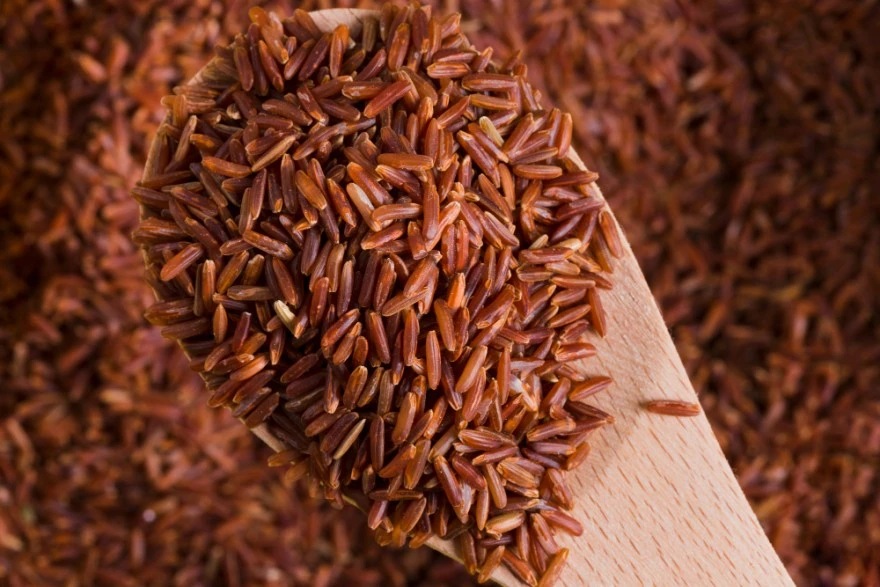
आरोग्यदायी आहारात तपकिरी तांदूळ असायलाच हवा याची 10 कारणे
तपकिरी तांदूळ म्हणजे काय? तपकिरी तांदूळ हे संपूर्ण धान्य आहे, ज्यामध्ये भुसी आणि गर्भाचे थर टिकून राहतात. पॉलिशिंग प्रक्रियेदरम्यान हे थर काढले जातात आणि त्यातून पांढरा तांदूळ तयार होतो. या थरांमध्ये तंतू, जीवनसत्त्वे, खनिजे आणि अँटिऑक्सिडंट्स असतात. या अल्प प्रक्रियेमुळे तपकिरी तांदळातील नैसर्गिक पोषणमूल्ये टिकून राहतात, ज्यामध्ये आवश्यक जीवनसत्त्वे, खनिजे आणि आहारातील तंतू यांचा समावेश होतो, त्यामुळे आरोग्याबाबत जागरूक व्यक्तींसाठी तो अधिक चांगला पर्याय ठरतो. तपकिरी तांदळाची खास खमंग चव आणि किंचित चिवट पोत हे त्याच्या अखंड राहिलेल्या बाहेरील थरांमुळे असते. तपकिरी तांदूळ आणि पांढरा तांदूळ यांची तुलना केली तर फरक लगेच लक्षात येतो—तपकिरी तांदूळ आपला नैसर्गिक रंग आणि पोषकतत्त्वांची घनता टिकवून ठेवतो, तर पांढऱ्या तांदळात पॉलिशिंगदरम्यान तंतू, मॅग्नेशियम आणि बी-जीवनसत्त्वे मोठ्या प्रमाणात कमी होतात. तपकिरी तांदळातील पोषकतत्त्वे: प्रत्येक दाण्यात काय असते? पोषकतत्त्व तपकिरी तांदूळ (1 कप, शिजवलेला) कॅलरीज 215–225 कार्बोहायड्रेट्स 45-52 g प्रथिने 4.5-5.5 g चरबी 1.6-2 g तंतू 3.2-3.5 g मॅग्नेशियम 84 mg (दैनंदिन गरजेच्या सुमारे 20%) मॅंगनीज 1.8 mg सेलेनियम 19 mcg तपकिरी तांदळाचे 10 फायदे जे दुर्लक्षित करता येणार नाहीत तंतूंचे भरपूर प्रमाण: तपकिरी तांदळात आहारातील तंतू मुबलक असतात, जे पचनक्रिया सुरळीत ठेवतात, बद्धकोष्ठतेपासून बचाव करतात आणि आतड्यांचे आरोग्य चांगले राखण्यास मदत करतात. मॅग्नेशियमचे उच्च प्रमाण: त्यातील लक्षणीय मॅग्नेशियम हाडे मजबूत करण्यास, मज्जासंस्थेची क्रिया समर्थित करण्यास आणि स्नायूंचे कार्य सुधारण्यास मदत करते. मॅंगनीजचा उत्कृष्ट स्रोत: तपकिरी तांदळातील मॅंगनीज चयापचय वाढवतो, अँटिऑक्सिडंट संरक्षण सुधारतो आणि कोलाजेन निर्मितीस मदत करतो. हृदयासाठी हितकारक गुणधर्म: नियमित सेवनामुळे कोलेस्टेरॉलची पातळी कमी होण्यास आणि रक्तदाब अधिक निरोगी राहण्यास मदत होऊ शकते, ज्यामुळे दीर्घकालीन हृदयआरोग्याला आधार मिळतो. रक्तातील साखरेचे नियमन: तपकिरी तांदळाचा मध्यम ग्लायसेमिक निर्देशांक (GI 50–55) रक्तातील ग्लुकोजची पातळी स्थिर ठेवण्यास मदत करतो आणि मधुमेह टाळण्यासही सहाय्यक ठरतो. वजन नियंत्रणास आधार: तपकिरी तांदळासारखी तंतुमय संपूर्ण धान्ये तृप्ती वाढवतात, भूक नियंत्रित ठेवतात आणि जास्त खाणे टाळण्यास मदत करतात. प्रतिकारशक्तीसाठी सेलेनियम: तपकिरी तांदळात सेलेनियम हे प्रभावी खनिज असते, जे प्रतिकारशक्ती मजबूत करण्यास आणि थायरॉईडचे संतुलन राखण्यास मदत करते. नैसर्गिकरित्या ग्लूटेन-मुक्त: हे धान्य नैसर्गिकरित्या ग्लूटेन-मुक्त असल्याने सीलिअॅक रोग किंवा ग्लूटेन संवेदनशीलता असलेल्या व्यक्तींसाठी उपयुक्त आहे. दीर्घकालीन आजारांपासून बचाव: यातील अँटिऑक्सिडंट्स आणि संपूर्ण धान्यातील पोषकतत्त्वे स्ट्रोक, काही प्रकारचे कर्करोग आणि इतर दीर्घकालीन आजारांचा धोका कमी करण्यास मदत करू शकतात. पाककलेतील बहुपयोगिता: तपकिरी तांदूळ अनेक पाककृतींमध्ये सहज वापरता येतो—बाउल्स, स्टिर-फ्रायपासून ते सॅलड्स आणि सूपपर्यंत—म्हणून आरोग्यदायी खाणे सोपे आणि आनंददायी बनते. आरोग्यदायी फायद्यांना चालना देणारी तपकिरी तांदळातील मुख्य पोषकतत्त्वे तपकिरी तांदळाचे उल्लेखनीय फायदे त्याच्या एकाग्र पोषकतत्त्वयुक्त रचनेतून मिळतात: आहारातील तंतू: पचनाचे आरोग्य आणि रक्तातील साखरेचे नियंत्रण सुधारतात मॅग्नेशियम: हाडांच्या आरोग्यासाठी आणि ऊर्जा निर्मितीसाठी आवश्यक मॅंगनीज: चयापचय आणि अँटिऑक्सिडंट संरक्षणास मदत करतो सेलेनियम: प्रतिकारशक्ती आणि डीएनए दुरुस्तीसाठी महत्त्वाचे बी-जीवनसत्त्वे: यामध्ये थायामिन, नायसिन आणि व्हिटॅमिन बी6 यांचा समावेश होतो फॉस्फरस: हाडांची निर्मिती आणि पेशींमधील ऊर्जेसाठी आवश्यक अँटिऑक्सिडंट संयुगे: पेशींच्या नुकसानीपासून संरक्षण करतात हृदयाच्या आरोग्यासाठी तपकिरी तांदळाचे फायदे तपकिरी तांदळातील पोषणमूल्ये अनेक मार्गांनी हृदयवाहिनीसंबंधी आरोग्यास महत्त्वाचे फायदे देतात: एलडीएल कोलेस्टेरॉल कमी करते: विद्रव्य तंतू कोलेस्टेरॉलचे कण बांधून शरीरातून बाहेर टाकण्यास मदत करतात रक्तदाब कमी करण्यास मदत: मॅग्नेशियम आणि पोटॅशियम निरोगी रक्ताभिसरणास आधार देतात धमनींमध्ये थर साचणे कमी करण्यास मदत: अँटिऑक्सिडंट्स रक्तवाहिन्यांची अखंडता जपतात हृदयाची लय समर्थित करते: आवश्यक खनिजे हृदयाचे कार्य योग्य प्रकारे चालू ठेवतात रक्तातील साखर आणि मधुमेह प्रतिबंधासाठी तपकिरी तांदळाचे फायदे मधुमेहासाठी तपकिरी तांदळाचे फायदे विशेषतः रक्तातील ग्लुकोज नियंत्रित ठेवणाऱ्यांसाठी उल्लेखनीय आहेत. अभ्यासांत असे दिसून आले आहे की पांढऱ्या तांदळाऐवजी तपकिरी तांदूळ घेतल्यास प्रकार 2 मधुमेहाचा धोका 23% पर्यंत कमी होऊ शकतो. कमी ग्लायसेमिक निर्देशांक: जेवणानंतर रक्तातील साखर झपाट्याने वाढणे टाळते सतत ऊर्जा मिळणे: गुंतागुंतीचे कार्बोहायड्रेट्स ग्लुकोज हळूहळू उपलब्ध करून देतात इन्सुलिन संवेदनशीलता सुधारते: तंतू शरीरातील ग्लुकोज प्रक्रियेस अधिक चांगला आधार देतात मधुमेहाचा धोका कमी: संपूर्ण धान्यांचे सेवन प्रकार 2 मधुमेहाचे प्रमाण कमी असण्याशी संबंधित आहे वजन नियंत्रणासाठी तपकिरी तांदळाचे फायदे आयसीएमआरच्या मते, आहाराची एकूण गुणवत्ता सुधारण्यासाठी आणि मधुमेह तसेच लठ्ठपणाचा धोका कमी करण्यासाठी पांढऱ्या तांदळासारख्या परिष्कृत पर्यायांपेक्षा तपकिरी तांदूळ आणि इतर संपूर्ण धान्ये निवडावीत. तपकिरी तांदूळ त्यातील तंतू आणि पोषकतत्त्वांच्या घनतेमुळे वजन नियंत्रणास मदत करतो, कारण तो भूक नियमन करण्यास आणि ऊर्जा संतुलन सुधारण्यास सहाय्यक ठरतो: तृप्ती अधिक काळ टिकते: उच्च तंतूमुळे जास्त वेळ पोट भरल्याची भावना राहते खाण्याची ओढ कमी होते: स्थिर रक्तातील साखर भुकेचे अचानक झटके टाळते चयापचयास आधार: बी-जीवनसत्त्वे ऊर्जा परिणामकारकपणे तयार होण्यासाठी मदत करतात प्रमाण नियंत्रण: पोषकतत्त्वांची घनता कमी प्रमाणातही पोषणाची गरज भागवते पचन आणि आतड्यांच्या आरोग्यासाठी तपकिरी तांदळाचे फायदे तपकिरी तांदळातील भरपूर तंतू पचनसंस्थेच्या आरोग्यात मोठा बदल घडवू शकतात. हे आहारातील तंतू प्रीबायोटिकसारखे काम करतात, उपयुक्त आतड्यांतील जिवाणूंना पोषण देतात आणि निरोगी मायक्रोबायोम वाढवतात. नियमित सेवनामुळे शौचक्रिया सुरळीत राहते, बद्धकोष्ठतेचा धोका कमी होतो आणि कोलोरेक्टल कर्करोगाचा धोका कमी असण्याशीही त्याचा संबंध आढळतो. याशिवाय, तपकिरी तांदळाचे फायदे असेही आहेत: पोषकतत्त्वांचे शोषण सुधारते पचनमार्गातील दाहसूचक घटक कमी होतात आतड्यांच्या संरक्षक भित्तीचे कार्य सुधारते पचनाचा एकूण आराम वाढतो तपकिरी तांदूळ विरुद्ध पांढरा तांदूळ: पोषणासाठी कोणता चांगला? वैशिष्ट्य तपकिरी तांदूळ पांढरा तांदूळ प्रक्रिया किमान (संपूर्ण धान्य) अधिक (परिष्कृत) तंतूंचे प्रमाण प्रति कप 3.5g प्रति कप 0.6g मॅग्नेशियम 84 mg 19 mg ग्लायसेमिक निर्देशांक कमी (50-55) जास्त (70-75) तृप्तीचा परिणाम जास्त कमी दीर्घकालीन आजारांपासून संरक्षण लक्षणीय अल्प ही तुलना स्पष्टपणे दाखवते की जवळजवळ प्रत्येक आरोग्यमानदंडात तपकिरी तांदळाचे पोषणमूल्य पांढऱ्या तांदळापेक्षा अधिक आहे. ग्लूटेन-मुक्त आणि अॅलर्जी-संवेदनशील आहारासाठी तपकिरी तांदळाचे फायदे तपकिरी तांदूळ नैसर्गिकरित्या ग्लूटेन-मुक्त आहे आणि सीलिअॅक रोग किंवा नॉन-सीलिअॅक ग्लूटेन संवेदनशीलता असलेल्या लोकांसाठी सुरक्षित आहे. गव्हावर आधारित धान्यांपेक्षा तपकिरी तांदूळ नैसर्गिकरित्या ग्लूटेन-मुक्त असूनही त्याचे पोषणमूल्य अधिक चांगले राहते. त्यामुळे मर्यादित आहार पाळणाऱ्या आणि तरीही पुरेशी प्रथिने व आवश्यक पोषकतत्त्वे हवी असलेल्या व्यक्तींसाठी तो विशेषतः उपयुक्त ठरतो. तपकिरी तांदळाबरोबर काळ्या तांदळाचे फायदे किंवा लाल तांदळाचे फायदे यांचाही समावेश केल्यास अँटिऑक्सिडंट्सने समृद्ध पोषणाचा आणखी एक थर मिळतो, ज्यामुळे विशिष्ट आहाराच्या गरजा असलेल्या व्यक्तींना अधिक पौष्टिक पर्याय मिळतात. दीर्घकालीन आजारांपासून बचावासाठी तपकिरी तांदळाचे फायदे तपकिरी तांदळाचे नियमित सेवन विविध आरोग्यधोक्यांना लक्षणीयरीत्या कमी करू शकते: प्रकार 2 मधुमेह प्रतिबंध: मेटा-विश्लेषणांनुसार संपूर्ण धान्यांचे सेवन मधुमेहाच्या सुमारे 16% कमी धोक्याशी संबंधित आहे हृदयविकाराचा धोका कमी होणे: कोलेस्टेरॉलचे प्रोफाइल आणि रक्तदाब सुधारतो कर्करोगापासून संरक्षण: अँटिऑक्सिडंट्स आणि तंतू कोलोरेक्टल कर्करोगाचा धोका कमी करण्यास मदत करू शकतात स्ट्रोक प्रतिबंध: रक्ताभिसरण सुधारते आणि दाहसूचक घटक कमी होतात मॅग्नेशियम, मॅंगनीज आणि सेलेनियममुळे तपकिरी तांदळाचे फायदे ही तीन खनिजे तपकिरी तांदळाच्या पोषणमूल्याला एकूण आरोग्यासाठी अत्यंत महत्त्वाचे बनवतात. मॅग्नेशियम 300 पेक्षा जास्त एन्झाइम प्रक्रियांना आधार देते, ज्यामध्ये ऊर्जा चयापचय आणि हाडांची निर्मिती यांचा समावेश होतो. मॅंगनीज प्रभावी अँटिऑक्सिडंट म्हणून काम करताना कार्बोहायड्रेट आणि अमिनो आम्लांच्या चयापचयास मदत करते. सेलेनियम प्रतिकारशक्ती सुधारते आणि डीएनए दुरुस्ती यंत्रणेमधील भूमिकेमुळे कर्करोगाचा धोका कमी करण्यास मदत करू शकते. जगभरातील पाककृतींमध्ये बहुपयोगी मुख्य अन्न म्हणून तपकिरी तांदूळ तपकिरी तांदूळ भारतीय पाककलेशी सहज जुळून जातो—सुगंधी बिर्याणीपासून आरामदायी डाळ-भाताच्या जोड्यांपर्यंत, तसेच खीर आणि पायसमसारख्या गोड पदार्थांपर्यंत. त्याचा ठसठशीत पोत मसालेदार करींबरोबर छान जुळतो, आणि त्याची खमंग चव पारंपरिक गोड पदार्थांची चवही अधिक खुलवते. भारतीय पाककलेपलीकडेही तपकिरी तांदूळ जागतिक पदार्थांमध्ये उत्तमरीत्या वापरता येतो—आशियाई स्टिर-फ्राय आणि थाई राईस बाउल्सपासून मेक्सिकन बुरिटोच्या सारणापर्यंत, भूमध्य शैलीतील तांदळाचे सॅलड्स आणि भरपूर पौष्टिक बुद्धा बाउल्सपर्यंत. पारंपरिक आणि आंतरराष्ट्रीय पाककृतींमध्ये सहज मिसळण्याची त्याची क्षमता त्याला चव किंवा सांस्कृतिक ओळख कमी न करता पौष्टिक आणि स्वादिष्ट पर्याय बनवते. संपूर्ण धान्याधारित आहारात शाश्वत पर्याय म्हणून तपकिरी तांदूळ तपकिरी तांदूळ निवडल्याने प्रक्रिया करण्यासाठी लागणारी ऊर्जा कमी होते आणि संपूर्ण धान्याधारित आहाराला प्रोत्साहन मिळते, त्यामुळे वैयक्तिक आरोग्याबरोबरच पर्यावरणीय शाश्वततेलाही आधार मिळतो. कमी प्रक्रियेमुळे पोषकतत्त्वे टिकून राहतात आणि ऊर्जा वापरही कमी होतो. संपूर्ण धान्यावर आधारित हा दृष्टिकोन व्यक्तीच्या आरोग्यासोबतच पृथ्वीच्या आरोग्यासाठीही उपयुक्त अशा शाश्वत खाण्याच्या पद्धतींशी सुसंगत आहे. चव आणि पोषकतत्त्वे उत्तम राहण्यासाठी तपकिरी तांदूळ कसा तयार करावा पाणी स्वच्छ येईपर्यंत थंड पाण्याखाली चांगले धुवा पचनास सोपा होण्यासाठी आणि शिजवण्याचा वेळ कमी करण्यासाठी 30-60 मिनिटे भिजत ठेवा योग्य पाण्याचे प्रमाण वापरा - सामान्यतः 2:1 पाणी ते तांदूळ उकळी आणा, नंतर झाकण ठेवून 40-50 मिनिटे मंद आचेवर शिजवा काट्याने हलके सैल करण्यापूर्वी 10 मिनिटे तसेच ठेवून द्या शिजवताना तमालपत्र किंवा अख्खे मसाले घाला शिजवलेला तपकिरी तांदूळ हवाबंद डब्यात भरून रेफ्रिजरेटरमध्ये 4–5 दिवसांपर्यंत ठेवा तपकिरी तांदळाचे संभाव्य तोटे आर्सेनिकचे जास्त प्रमाण: विशेषतः काही प्रदेशांत पिकवलेल्या तपकिरी तांदळामध्ये अकार्बनिक आर्सेनिकचे प्रमाण किंचित जास्त असू शकते. विविध धान्यांचे सेवन करणे आणि तांदूळ जास्त पाण्यात शिजवणे यामुळे संपर्क कमी करण्यास मदत होऊ शकते. शिजवण्यास अधिक वेळ: पांढऱ्या तांदळापेक्षा याला साधारणपणे अधिक वेळ भिजवणे आणि जास्त वेळ शिजवणे लागते, त्यामुळे अतिरिक्त नियोजन आणि तयारीची गरज पडू शकते. पचनाशी जुळवून घेणे: उच्च तंतूमुळे तंतुमय अन्नाची सवय नसलेल्या व्यक्तींमध्ये सुरुवातीला पोट फुगणे किंवा वायू होऊ शकतो, मात्र काळानुसार हे अनेकदा सुधारते. फायटिक आम्लाची उपस्थिती: तपकिरी तांदळात फायटिक आम्ल हे नैसर्गिक अँटिऑक्सिडंट असते, जे खनिजांचे शोषण किंचित कमी करू शकते; शिजवण्यापूर्वी तांदूळ भिजवणे किंवा आंबवणे यामुळे हा परिणाम कमी करण्यास मदत होते. साठवणुकीची काळजी: नैसर्गिक तेलांमुळे तपकिरी तांदळाची साठवणकालमर्यादा कमी असते आणि थंड, हवाबंद जागेत योग्य प्रकारे न ठेवल्यास तो लवकर खराब होऊ शकतो. निष्कर्ष तपकिरी तांदूळ हे असे पोषणसमृद्ध अन्न आहे, जे साध्या जेवणालाही आरोग्यवर्धक अनुभवात बदलू शकते. रक्तातील साखरेचे नियंत्रण, हृदयाच्या आरोग्यास आधार, पचनक्रिया सुधारणा आणि दीर्घकालीन आजारांपासून बचाव—दैनंदिन आहारात तपकिरी तांदूळ समाविष्ट करण्याचे फायदे तात्काळही जाणवू शकतात आणि दीर्घकाळ टिकणारेही असतात. मेट्रोपोलिस हेल्थकेअरमध्ये आम्ही तुमच्या आरोग्यप्रवासाला सर्वसमावेशक निदान सेवांद्वारे साथ देतो, ज्यामुळे आहारातील बदलांचा—उदा. जेवणात तपकिरी तांदूळ समाविष्ट करण्याचा—सकारात्मक परिणाम लक्षात ठेवता येतो. मधुमेह पॅनल्स, लिपिड प्रोफाइल्स आणि पोषणमूल्य चाचण्यांसह 4,000 पेक्षा अधिक चाचण्यांचे आमचे विस्तृत जाळे तुम्हाला तुमचे आरोग्य अधिक चांगले राखण्यासाठी आवश्यक माहिती देते. 98% पेक्षा जास्त सीएपी प्रावीण्य गुणांसह, ज्यामुळे जागतिक स्तरावरील सर्वोच्च 1% प्रयोगशाळांमध्ये त्याची गणना होते, मेट्रोपोलिस अचूक आणि विश्वासार्ह निष्कर्ष सुनिश्चित करते. वारंवार विचारले जाणारे प्रश्न तपकिरी तांदूळ खरोखर पांढऱ्या तांदळापेक्षा अधिक आरोग्यदायी आहे का? नक्कीच. तपकिरी तांदळाच्या फायद्यांमध्ये पांढऱ्या तांदळाच्या तुलनेत लक्षणीयरीत्या जास्त तंतू, मॅग्नेशियम आणि अँटिऑक्सिडंट्स यांचा समावेश होतो. त्याची संपूर्ण धान्यरचना टिकाऊ ऊर्जा आणि रक्तातील साखरेचे अधिक चांगले नियंत्रण देते, त्यामुळे बहुतेक व्यक्तींसाठी तो पोषणदृष्ट्या अधिक चांगला पर्याय ठरतो. वजन कमी करण्यासाठी तपकिरी तांदळाचे मुख्य फायदे कोणते? तपकिरी तांदळातील कॅलरीज वजन नियंत्रणासाठी प्रभावी ठरतात, कारण तो तृप्ती वाढवतो, रक्तातील साखरेची पातळी स्थिर ठेवतो आणि चयापचय सुधारतो. त्यातील जास्त तंतू तुम्हाला जास्त वेळ पोटभरल्यासारखे वाटू देतात, त्यामुळे नैसर्गिकरीत्या एकूण कॅलरी सेवन कमी होते आणि आवश्यक पोषकतत्त्वेही मिळतात. मधुमेह असलेल्या लोकांसाठी तपकिरी तांदूळ योग्य आहे का? होय, मधुमेहासाठी तपकिरी तांदूळ अनेक फायदे देतो. मधुमेहासाठी तपकिरी तांदळाचे फायदे यामध्ये परिष्कृत धान्यांच्या तुलनेत कमी ग्लायसेमिक निर्देशांक, रक्तातील साखरेचे अधिक चांगले नियंत्रण आणि इन्सुलिन संवेदनशीलतेत सुधारणा यांचा समावेश होतो. मात्र, उत्तम ग्लुकोज नियंत्रणासाठी प्रमाणात खाणे महत्त्वाचे राहते. दररोज किती तपकिरी तांदूळ खावा? अर्धा कप शिजवलेल्या तपकिरी तांदळाचे प्रमाण जास्त कॅलरीज न घेता उत्तम पोषण देते. या प्रमाणातून सुमारे 108 कॅलरीज आणि भरपूर पोषकतत्त्वे मिळतात, तसेच निरोगी वजन नियंत्रण आणि रक्तातील साखरेच्या नियंत्रणालाही आधार मिळतो. तपकिरी तांदूळ दररोज खाणे सुरक्षित आहे का? दररोज तपकिरी तांदूळ खाणे सर्वसाधारणपणे सुरक्षित आणि फायदेशीर आहे. मात्र, आर्सेनिकचा संपर्क कमी ठेवण्यासाठी आणि आहारात विविधता राखण्यासाठी वेगवेगळी धान्ये वापरा. उत्तम पोषणासाठी तपकिरी तांदळाबरोबर क्विनोआ, बाजरी आणि ओट्ससारखी इतर संपूर्ण धान्येही समाविष्ट करा. तपकिरी तांदळात आर्सेनिक असते का? धोका कसा कमी करावा? तपकिरी तांदळात नैसर्गिकरित्या आर्सेनिक असते, जरी त्याचे प्रमाण पिकवलेल्या प्रदेशानुसार बदलते. तांदूळ नीट धुणे, जास्त पाण्यात शिजवणे आणि वेगवेगळी धान्ये वापरणे यामुळे संपर्क कमी करता येतो. प्रमाणात सेवन केल्यास त्याचे आरोग्यदायी फायदे सहसा धोक्यांपेक्षा जास्त ठरतात.
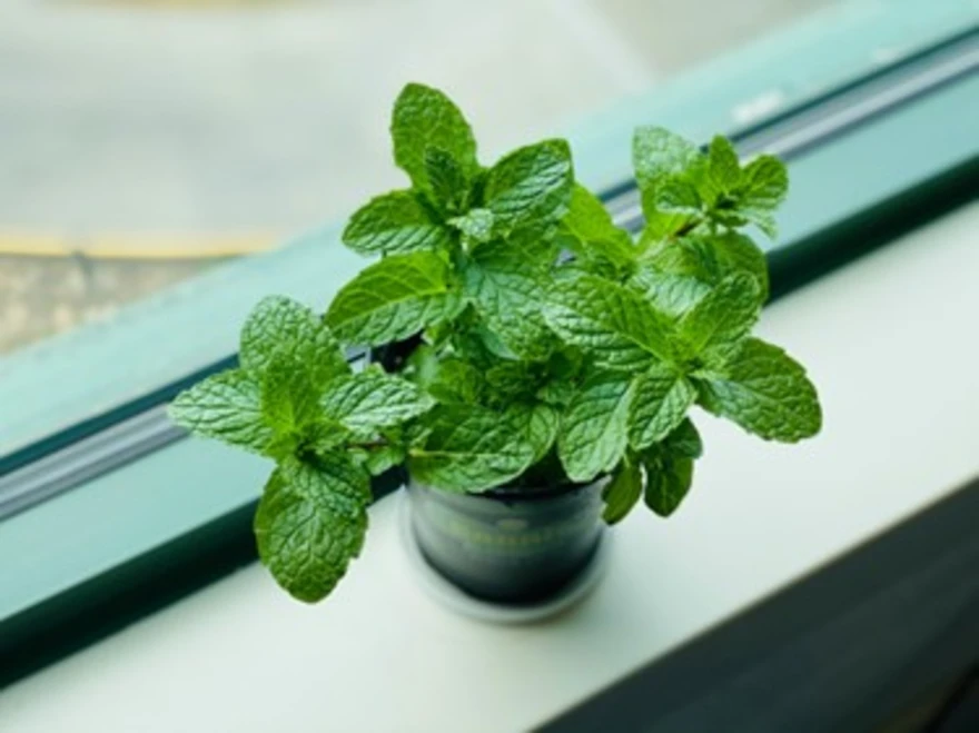
पुदिन्याचे फायदे: पचन, त्वचा आणि ताज्या श्वासासाठी या शीतल औषधी वनस्पतीचे 9 आश्चर्यकारक उपयोग
पुदीना म्हणजे काय? पुदीना (जीनस मेंथा) ही लॅमिएसी कुलातील एक बहुवर्षायू औषधी वनस्पती आहे. यामध्ये पेपरमिंट, स्पिअरमिंट आणि रानपुदिना यांसह 20 पेक्षा अधिक ज्ञात प्रजातींचा समावेश होतो. मूळची युरोप आणि आशियातील असलेली ही वनस्पती आता तिच्या औषधी आणि पाककलेतील उपयोगामुळे जगभर लागवड केली जाते. पुदिन्याचा वैशिष्ट्यपूर्ण सुगंध मेंटॉलमुळे असतो. हे एक नैसर्गिक संयुग आहे, जे थंडावा देणारी जाणीव निर्माण करते आणि कोंदलेपणा कमी करण्यास मदत करते. याची कोवळी हिरवी पाने ताजी किंवा वाळवून पेये, कोशिंबिरी, सॉस आणि डेझर्ट्सना चव देण्यासाठी वापरली जातात. आयुर्वेदानुसार, पुदिन्याला ‘शीतल’ औषधी वनस्पती मानले जाते, जी पित्त दोष संतुलित ठेवण्यास आणि निरोगी पचनाग्नीला आधार देण्यास मदत करते. पुदिन्याची पाने, पुदिन्याचे पाणी किंवा आवश्यक तेल अशा कोणत्याही स्वरूपात असो, ही साधी वनस्पती नैसर्गिक उपचारपद्धती आणि आधुनिक पोषणशास्त्रात आजही महत्त्वाची भूमिका बजावत आहे. पुदिन्याचे पोषणमूल्य पुदिन्याची पाने कमी प्रमाणात वापरली जात असली तरी ती आवश्यक पोषक घटकांनी आश्चर्यकारकरित्या समृद्ध असतात. ताज्या पुदिन्याच्या 100-ग्राम प्रमाणात काय मिळते ते येथे दिले आहे (अंदाजे मूल्ये): कॅलरी: 44 kcal पाणी: 85% प्रथिने: 3.3 g आहारातील तंतू: 8 g व्हिटॅमिन A: डेली व्हॅल्यू (DV) च्या 23%, दृष्टी आणि त्वचेच्या आरोग्यास आधार देते व्हिटॅमिन C: DV च्या 20%, रोगप्रतिकारक शक्ती आणि कोलेजन निर्मितीस आधार देते लोह: DV च्या 16%, लाल रक्तपेशी निर्मितीसाठी महत्त्वाचे फोलेट: DV च्या 6%, पेशींची वाढ आणि चयापचयास आधार देते कॅल्शियम: DV च्या 20%, हाडे आणि दात मजबूत करते अँटिऑक्सिडंट्स: फ्लॅव्होनॉइड्स, रोझमॅरिनिक अॅसिड आणि मेंटॉल, जे पेशींना ऑक्सिडेटिव्ह ताणापासून संरक्षण देण्यास मदत करतात पोषक घटक आणि जैवसक्रिय संयुगांच्या या मिश्रणामुळे, दैनंदिन आहारात अगदी कमी प्रमाणात पुदिन्याची पाने घातली तरी पचन, रोगप्रतिकारक शक्ती आणि ताजेतवानेपणा यांना लक्षणीय मदत मिळू शकते. पुदिन्याचे 9 आरोग्यदायी फायदे: ही औषधी वनस्पती आरोग्यासाठी इतकी प्रभावी का आहे 1. पचनास मदत करते आणि पोटफुगी कमी करते पुदिन्यातील मेंटॉल पचनमार्गातील सपाट स्नायूंना शिथिल करते, ज्यामुळे अन्न सहज पुढे सरकते आणि वायू किंवा पोटफुगीपासून आराम मिळतो. अभ्यास दर्शवतात की पेपरमिंट तेल इरिटेबल बाउल सिंड्रोम (IBS)ची लक्षणे कमी करू शकते, कारण ते आतड्यांतील आकडी शांत करण्यास मदत करते. 2. नैसर्गिकरित्या श्वास ताजा ठेवते पुदिन्याचे जिवाणूरोधक गुणधर्म तोंडातील हानिकारक जिवाणूंना आळा घालतात आणि दीर्घकाळ टिकणारा थंडावा देतात. पुदिन्याची पाने चावणे किंवा पुदिन्याचा चहा पिणे यामुळे दुर्गंधी कमी होण्यास आणि तोंडाची स्वच्छता राखण्यास मदत होते. 3. अपचन आणि छातीत जळजळ कमी करते जेवणानंतर पुदिन्याचे पाणी किंवा पुदिन्याचा चहा घेतल्याने पित्तस्राव वाढण्यास आणि तेलकट अन्नाचे पचन होण्यास मदत मिळू शकते, ज्यामुळे जेवणानंतरचा त्रास कमी होतो. 4. श्वसनाच्या आरोग्यास आधार देते पुदिन्यातील मेंटॉल नैसर्गिक कफनाशकासारखे कार्य करते. ते नाकाचे मार्ग मोकळे करण्यास, घशातील जळजळ कमी करण्यास आणि सामान्य सर्दी किंवा सायनुसायटिसची लक्षणे कमी करण्यास मदत करते. पुदिन्याच्या वाफेचा श्वास घेतल्याने झटपट आराम मिळू शकतो. 5. निरोगी त्वचेला प्रोत्साहन देते पुदिन्याचे जिवाणूरोधक आणि दाहरोधक परिणाम मुरुमांशी लढण्यास, जळजळ शांत करण्यास आणि निस्तेज त्वचेला उजळ करण्यास मदत करतात. घरच्या घरी तयार केलेले पुदिन्याचे फेस पॅक किंवा टोनर लालसरपणा कमी करू शकतात आणि त्वचेची रंध्रे स्वच्छ ठेवू शकतात. 6. डोकेदुखी आणि ताण कमी करते कपाळाच्या दोन्ही बाजूंना पातळ केलेले पेपरमिंट तेल लावल्याने किंवा पुदिन्याच्या वाफेचा श्वास घेतल्याने स्नायू शिथिल होतात आणि तणावामुळे होणाऱ्या डोकेदुखीपासून आराम मिळतो. त्याचा सुगंध स्वतःच शांतता आणि मानसिक स्पष्टतेची भावना निर्माण करतो. 7. वजन नियंत्रणास मदत करते पुदिन्याचा चहा आणि पुदिन्याचे पाणी पचन सुधारतात, खाण्याची अनावश्यक इच्छा कमी करतात आणि चयापचयाला आधार देतात. पुदिन्याचे पाणी पिऊन शरीरात पाणी टिकवून ठेवल्यास ताजेतवाने वाटते आणि कॅलरीचे सेवन कमी करण्यास मदत मिळू शकते. 8. मेंदूची कार्यक्षमता आणि लक्ष वाढवते संशोधन असे सूचित करते की पुदिन्याचा वास घेणे किंवा त्याचे सेवन करणे यामुळे मध्यवर्ती मज्जासंस्थेला चालना मिळून जागरूकता, स्मरणशक्ती आणि एकाग्रता वाढू शकते. 9. रोगप्रतिकारक शक्ती आणि शरीरशुद्धीला आधार देते पुदिना अँटिऑक्सिडंट्सने समृद्ध असतो, जे पेशींना फ्री रॅडिकल्सपासून संरक्षण देतात, रोगप्रतिकारक प्रतिसाद बळकट करतात आणि शरीराच्या नैसर्गिक विषद्रव्ये बाहेर टाकण्याच्या प्रक्रियेला मदत करतात. जास्तीत जास्त फायद्यासाठी पुदिना कसा वापरावा पुदिना आपल्या दिनचर्येत समाविष्ट करणे सोपे आणि बहुपयोगी आहे. तो दैनंदिन आयुष्यात वापरण्याचे काही सोपे आणि परिणामकारक मार्ग येथे दिले आहेत: पुदिन्याचा चहा: ताजी किंवा वाळवलेली पुदिन्याची पाने गरम पाण्यात 5–7 मिनिटे मुरू द्या. हा हर्बल चहा पचन शांत करतो, पोटफुगी कमी करतो आणि मन ताजेतवाने करतो. पुदिन्याचे पाणी: पुदिन्याची पाने, लिंबाचे काप किंवा काकडी घालून पाणी रात्रभर मुरू द्या. यामुळे शरीरशुद्धीला मदत करणारे आणि शरीराला पाणीपुरवठा करणारे पेय तयार होते. पाककलेतील उपयोग: तात्काळ ताजेपणा आणि छान चव मिळण्यासाठी चिरलेली पुदिन्याची पाने कोशिंबीर, चटणी, सूप किंवा दह्यात घाला. पुदिन्याचे तेल: डोकेदुखी आणि स्नायूंचा ताण कमी करण्यासाठी अरोमाथेरपीमध्ये किंवा बाहेरून लावण्यासाठी पातळ केलेले पेपरमिंट तेल वापरा. त्वचेची काळजी: मुरुमे आणि दाह शांत करण्यासाठी कुटलेली पुदिन्याची पाने गुलाबपाणी किंवा कोरफडीसोबत मिसळून थंडावा देणारा फेस पॅक तयार करा. सर्दीसाठी घरगुती उपाय: नाकातील कोंदलेपणा आणि सायनस दाब कमी करण्यासाठी पुदिन्याची पाने घातलेल्या वाफेचा श्वास घ्या. या सोप्या पद्धतींमध्ये पारंपरिक आयुर्वेदीय पद्धती आणि आधुनिक आरोग्यदायी सवयी दोन्हींचा समावेश आहे, ज्यामुळे वर्षभर पुदिन्याचे फायदे घेता येतात. पुदिन्याच्या फायद्यांवरील वैज्ञानिक अभ्यास काही संशोधन अभ्यासांनी पुदिन्याचे शारीरिक आणि उपचारात्मक गुणधर्म तपासले आहेत: जर्नल ऑफ क्लिनिकल गॅस्ट्रोएन्टेरोलॉजी (2014) मध्ये प्रकाशित झालेल्या मेटा-विश्लेषणात असे आढळले की पेपरमिंट तेलाच्या कॅप्सूल्स आयबीएसची लक्षणे आणि पोटदुखी सुधारण्यात प्लेसिबोपेक्षा लक्षणीयरीत्या अधिक प्रभावी होत्या, ज्यामुळे त्यांची अल्पकालीन सुरक्षितता आणि परिणामकारकता सिद्ध झाली. एनआयएचचे मेंथा पाइपेरिटा (पेपरमिंट) वरील संशोधन तोंडातील रोगकारक जिवाणूंविरुद्ध, जसे स्ट्रेप्टोकॉकस म्युटन्स आणि कँडिडा अल्बिकन्स, प्रभावी सूक्ष्मजंतुरोधक आणि अँटी-बायोफिल्म गुणधर्म दर्शवते. अभ्यास सूचित करतात की पेपरमिंट तेल आणि पानांचे अर्क, विशेषतः काइटोसन नॅनो-जेल्समध्ये तयार केल्यावर, दातांवरील प्लॅक आणि हिरड्यांतील दाह प्रभावीपणे कमी करू शकतात, ज्यामुळे सुधारित तोंडाच्या स्वच्छतेसाठी टूथपेस्ट आणि माउथवॉशमध्ये त्यांचा वापर समर्थित होतो. ह्यूमन सायकोफार्माकोलॉजीमध्ये प्रकाशित 2023 च्या यादृच्छिक प्लेसिबो-नियंत्रित चाचणीत असे आढळले की 200 mL पेपरमिंट चहा पिल्याने स्मरणशक्ती, लक्ष आणि कार्यरत स्मरणशक्तीमध्ये लक्षणीय सुधारणा झाली, तसेच प्रीफ्रंटल कॉर्टेक्समधील मेंदूकडे जाणारा रक्तप्रवाह वाढला. या अभ्यासाने निष्कर्ष काढला की पेपरमिंट संज्ञानात्मक कार्यक्षमता आणि मेंदूतील प्राणवायू पुरवठा सुधारतो, ज्यामुळे निरोगी प्रौढांसाठी नैसर्गिक नूट्रोपिक म्हणून त्याची क्षमता अधोरेखित होते. हे निष्कर्ष एकत्रितपणे आयुर्वेदासारख्या पारंपरिक प्रणालींना पूर्वीपासून माहीत असलेली गोष्ट सिद्ध करतात: पुदिन्याची पाने पचनास मदत करतात, रोगप्रतिकारक शक्ती वाढवतात आणि एकूणच आरोग्य सुधारतात. पुदिन्याचे संभाव्य दुष्परिणाम आणि घ्यावयाची खबरदारी पुदिना सामान्यतः मर्यादित प्रमाणात सेवन केल्यास सुरक्षित असतो, परंतु अतिवापर किंवा काही विशिष्ट आरोग्यस्थितींमध्ये दुष्परिणाम होऊ शकतात. लक्षात ठेवण्यासारख्या काही खबरदारी पुढीलप्रमाणे आहेत: अॅसिड रिफ्लक्स (GERD): पुदिना अन्ननलिकेच्या तळाशी असलेला स्नायू शिथिल करू शकतो, ज्यामुळे काही वेळा अॅसिड रिफ्लक्स किंवा छातीत जळजळ वाढू शकते. अॅलर्जी: मेंटॉल किंवा लॅमिएसी कुलातील वनस्पतींना (जसे तुळस किंवा सेज) अॅलर्जी असलेल्या लोकांना सौम्य चिडचिड किंवा त्वचेवर पुरळ येऊ शकते. गर्भधारणा: गर्भधारणेदरम्यान एकाग्र पुदिन्याचे तेल किंवा अतितीव्र पेपरमिंट चहा जास्त प्रमाणात घेणे शिफारसीय नाही, कारण त्याचे गर्भाशय शिथिल करणारे परिणाम असू शकतात. अर्भक आणि लहान मुले: अर्भकांच्या चेहऱ्यावर किंवा छातीवर मेंटॉल तेल थेट लावू नका, कारण त्यामुळे श्वासोच्छ्वासाच्या अडचणी निर्माण होऊ शकतात. औषधांशी परस्परसंवाद: पुदिन्याच्या तेलाचे जास्त डोस रक्तदाब किंवा मधुमेहासाठीच्या काही औषधांमध्ये अडथळा आणू शकतात. आवश्यक तेलं: त्वचेची जळजळ किंवा आग होण्यासारखी जाणीव टाळण्यासाठी बाहेरून वापरण्यापूर्वी पुदिन्याचे तेल नेहमी पातळ करा. सामान्यतः, अन्नात किंवा चहात वापरली जाणारी ताजी पुदिन्याची पाने बहुतेक लोकांसाठी सुरक्षित आणि फायदेशीर असतात. पुदिना सर्वांसाठी सुरक्षित आहे का? बहुतेक निरोगी प्रौढांसाठी, पुदिन्याची पाने किंवा पुदिन्याचे पाणी रोज घेणे सुरक्षित आणि पोषक असते. मात्र, अॅसिड रिफ्लक्स, पित्ताशयाचे आजार किंवा ज्ञात अॅलर्जी असलेल्या व्यक्तींनी ते सावधगिरीने वापरावे. मुलांनी आणि गर्भवती महिलांनी एकाग्र पुदिन्याचे तेल किंवा पूरक वापरण्यापूर्वी डॉक्टरांचा सल्ला घ्यावा. मर्यादित प्रमाण महत्त्वाचे आहे: आपल्या जेवणात काही ताजी पाने किंवा दिवसातून एक कप पुदिन्याचा चहा इतकाच त्याच्या नैसर्गिक थंडावा देणाऱ्या आणि शुद्धीकारक गुणांचा लाभ घेण्यासाठी पुरेसा आहे. निष्कर्ष ताजा, सुगंधी आणि उपचारक्षमतेने समृद्ध असा पुदिना पचनाच्या आरोग्यास, स्वच्छ त्वचेला आणि संतुलित रोगप्रतिकारक शक्तीला आधार देतो. प्राचीन आयुर्वेद आणि आधुनिक विज्ञान या दोन्हींचा आधार असलेली ही बहुपयोगी औषधी वनस्पती पुदिन्याचा चहा, पुदिन्याचे पाणी किंवा जेवणातील ताजी पाने अशा कुठल्याही स्वरूपात आपल्या दैनंदिन आरोग्य दिनचर्येत साधी पण प्रभावी भर ठरते. Metropolis Healthcareमुळे आपल्या आरोग्याबाबत सजग राहणे अधिक सोपे होते. 4000+ निदान चाचण्या, सर्वसमावेशक संपूर्ण शरीर तपासण्या आणि विशेष चाचण्यांसह आम्ही अचूक माहिती आणि मनःशांती देतो. 10,000+ ठिकाणांवरून घरपोच नमुना संकलन, जलद आणि अचूक अहवाल, तसेच आमची वेबसाइट, अॅप, व्हॉट्सअॅप किंवा फोनद्वारे बुकिंगची सुविधा यांचा लाभ घ्या. NABL & CAP मान्यताप्राप्त प्रयोगशाळांच्या आधारावर, Metropolis विश्वासार्ह निदान क्षेत्रात नवे मानदंड प्रस्थापित करत आहे — जे आपल्याला अधिक चांगल्या आरोग्याकडे आत्मविश्वासाने प्रत्येक पाऊल टाकण्यास मदत करते. सामान्य प्रश्न दररोज पुदिन्याची पाने खाल्ल्याने कोणते फायदे होतात? दररोज पुदिन्याची पाने खाल्ल्याने पचनास मदत होते, श्वास ताजा राहतो, रोगप्रतिकारक शक्ती वाढते आणि दाह व संसर्गांपासून संरक्षण देणारे अँटिऑक्सिडंट्स मिळतात. पुदिना वजन कमी करण्यास मदत करू शकतो का? होय. पुदिन्याचे पाणी किंवा पुदिन्याचा चहा पचनास मदत करतो, चयापचय सुधारतो आणि खाण्याची अनावश्यक इच्छा कमी करण्यास मदत करतो — आणि हे सर्व अप्रत्यक्षपणे निरोगी वजन नियंत्रणास आधार देतात. पचनासाठी पुदिन्याचा चहा किती वेळा घ्यावा? पोटफुगी कमी करण्यासाठी आणि पचन सुरळीत ठेवण्यासाठी दिवसातून एक किंवा दोन कप पुदिन्याचा चहा, शक्यतो जेवणानंतर, योग्य मानला जातो. पुदिन्याचा वापर त्वचेच्या काळजीसाठी करता येतो का? नक्कीच. पुदिन्याचे जिवाणूरोधक आणि थंडावा देणारे गुणधर्म मुरुमे नियंत्रणात ठेवण्यास, जळजळ कमी करण्यास आणि मास्क किंवा टोनरमध्ये वापरल्यास त्वचेला ताजेतवाने चमक देण्यास मदत करतात. ताजा पुदिना किंवा पुदिन्याचे तेल कुठे खरेदी करता येते? ताजी पुदिन्याची पाने सुपरमार्केट, स्थानिक बाजार आणि ऑनलाइन किराणा दुकानांमध्ये सहज उपलब्ध असतात. शुद्ध पुदिन्याचे आवश्यक तेल नामांकित आयुर्वेदीय किंवा सेंद्रिय ब्रँड्सकडून खरेदी करता येते.
ब्राझिल नट्सचे 5 अविश्वसनीय आरोग्यदायी फायदे, जे तुम्हाला माहित नव्हते
ब्राझिल नट्स म्हणजे काय? ब्राझिल नट्स या मोठ्या, त्रिकोणी बिया आहेत ज्या ब्राझिल नटच्या झाडापासून (बर्थोलेटिया एक्सेल्सा) मिळतात. ही ॲमेझॉन प्रदेशातील मूळ प्रजाती असून 50 metres पर्यंत उंच वाढू शकते आणि शतकानुशतके जगू शकते; काही झाडे 500 वर्षांहून अधिक जगतात. ही प्रभावी झाडे ब्राझिल, बोलिव्हिया आणि पेरू येथील स्वच्छ, नैसर्गिक ॲमेझॉन वर्षावनातच चांगली वाढतात. ब्राझिल नटची झाडे परागीभवनासाठी वर्षावनातील विशिष्ट परिस्थिती आणि मोठ्या मधमाशींच्या प्रजातींवर (युग्लोसा spp.) अवलंबून असतात, त्यामुळे मोठ्या प्रमाणावर लागवड करणे अत्यंत कठीण ठरते. ब्राझिल नटच्या प्रत्येक शेंगामध्ये 12-24 स्वतंत्र नट्स असतात, जे संत्र्याच्या फोडींप्रमाणे मांडलेले असतात. या नट्सची चव समृद्ध, बटरसारखी आणि किंचित गोडसर, मातीसारखी असते, त्यामुळे ते अतिशय समाधानकारक वाटतात. ब्राझिल नट्सना इतर नट्सपासून वेगळे ठरवणारी गोष्ट म्हणजे त्यातील अपवादात्मक सेलेनियमचे प्रमाण; फक्त एक ब्राझिल नट तुमच्या रोजच्या सेलेनियम गरजेची पूर्तता करू शकतो किंवा ती ओलांडू शकतो. ब्राझिल नट्सचे पोषणमूल्य ब्राझिल नट्स हे पोषणमूल्यांनी समृद्ध अन्नपदार्थ का आहेत, हे समजून घेतल्यास त्यांचे उल्लेखनीय आरोग्यदायी फायदे स्पष्ट होतात: सेलेनियमचा उत्कृष्ट स्रोत: सेलेनियमचे प्रमाण प्रत्येक नटामध्ये 50–100 µg इतके बदलू शकते (रोजच्या गरजेच्या 90–180%), आणि ते ज्या जमिनीत वाढते त्यावर अवलंबून असते. आरोग्यदायी चरबी: हृदयवहिन्यासंबंधी आरोग्यास मदत करणाऱ्या मोनोअनसॅच्युरेटेड आणि पॉलीअनसॅच्युरेटेड चरबींनी समृद्ध उत्तम प्रथिने: 28-gram प्रमाणात सुमारे 4 grams आवश्यक खनिजे: मॅग्नेशियम (रोजच्या गरजेच्या 26%), कॉपर, मॅंगनीज, फॉस्फरस आणि झिंकचे भरपूर प्रमाण व्हिटॅमिन्स: थायमिन (व्हिटॅमिन B1) आणि व्हिटॅमिन E चा चांगला स्रोत तंतूंचे प्रमाण: पचनाच्या आरोग्यासाठी आहारातील तंतू पुरवते कमी कर्बोदके: नैसर्गिकरीत्या कमी carbs आणि sodium-free ब्राझिल नट्सचे फायदे सेलेनियमपासूनच कसे सुरू होतात ब्राझिल नट्सचे असामान्य फायदे प्रामुख्याने त्यांच्या अतुलनीय सेलेनियमच्या प्रमाणामुळे आहेत. सेलेनियम हे एक अत्यावश्यक सूक्ष्म खनिज आहे, जे उत्तम आरोग्यासाठी शरीराला आवश्यक असते, पण शरीर ते स्वतः तयार करू शकत नाही. हे प्रभावी अँटिऑक्सिडंट खनिज सेलेनोप्रोटीन्सच्या निर्मितीस मदत करते, जी डीएनए निर्मिती, थायरॉईड हार्मोन चयापचय आणि पेशींचे संरक्षण यामध्ये महत्त्वाची भूमिका बजावतात. क्लिनिकल अभ्यासांनुसार, सेलेनियमची कमतरता असलेल्या प्रदेशांमध्ये अनेक आठवडे रोज एक ब्राझिल नट खाल्ल्यास सेलेनियमची पातळी सामान्य होऊ शकते. द जर्नल ऑफ न्यूट्रिशन अँड मेटाबॉलिझमच्या मते, त्यामुळे ते जगभरातील सेलेनियमच्या सर्वात समृद्ध अन्नस्रोतांपैकी एक ठरतात. मात्र, ब्राझिल नटचे झाड ज्या जमिनीत वाढते त्या जमिनीच्या परिस्थितीनुसार सेलेनियमचे प्रमाण लक्षणीयरीत्या बदलते. काही नट्समध्ये प्रत्येक नटामागे 91 mcg पर्यंत सेलेनियम असू शकते, तर काहींमध्ये त्यापेक्षा खूप कमी असू शकते. ब्राझिल नट्सचे 5 अविश्वसनीय आरोग्यदायी फायदे 1. प्रभावी अँटिऑक्सिडंट संरक्षण ब्राझिल नट्समधील सेलेनियम शरीरातील नैसर्गिक अँटिऑक्सिडंट एन्झाइम्सची क्रिया वाढवून उत्कृष्ट अँटिऑक्सिडंट संरक्षण देते. हे एन्झाइम्स मुक्त रॅडिकल्समुळे होणाऱ्या ऑक्सिडेटिव्ह हानीपासून पेशींचे संरक्षण करतात, ज्यामुळे हृदयविकार आणि काही प्रकारच्या कर्करोगांसह दीर्घकालीन आजारांचा धोका कमी होऊ शकतो. मर्यादित प्रमाणात नियमित सेवन केल्यास, ते ऑक्सिडेटिव्ह ताण आणि दाहाविरुद्ध शरीराच्या संरक्षणाला मदत करू शकते. 2. थायरॉईडच्या कार्याला मदत करते थायरॉईड ग्रंथीला निष्क्रिय थायरॉईड हार्मोन्सना सक्रिय स्वरूपात रूपांतरित करण्यासाठी सेलेनियमची आवश्यकता असते. थायरॉईडच्या आरोग्यासाठी ब्राझिल नट्सचे फायदे विशेषतः महत्त्वाचे आहेत—पुरेसे सेलेनियम सेवन थायरॉईडचे निरोगी कार्य राखण्यास मदत करते आणि थायरॉईड विकार टाळण्यास सहाय्य करू शकते. योग्य चयापचय, ऊर्जा पातळी आणि शरीराचे तापमान नियंत्रण राखण्यासाठी हे विशेषतः महत्त्वाचे आहे. 3. हृदयाचे आरोग्य सुधारते ब्राझिल नट्समधील आरोग्यदायी असंतृप्त चरबी हानिकारक एलडीएल कोलेस्टेरॉल कमी करून आणि उपयुक्त एचडीएल कोलेस्टेरॉल वाढवून तुमचे कोलेस्टेरॉल प्रोफाइल सुधारण्यास मदत करते. अभ्यास सुचवतात की मर्यादित सेवनामुळे कोलेस्टेरॉल प्रोफाइल सुधारू शकते आणि रक्तातील लिपिड पातळ्या सुधारून हृदयवहिन्यासंबंधी आरोग्यास मदत होऊ शकते, जरी दीर्घकालीन परिणामांवरील माहिती मर्यादित आहे. 4. मेंदूचे कार्य आणि मनःस्थिती सुधारते सेलेनियम मेंदूच्या आरोग्यात आणि मनःस्थितीच्या नियमनात महत्त्वाची भूमिका बजावते. पुरेसे सेलेनियम सेवन सुधारलेल्या मनःस्थितीशी आणि संज्ञानात्मक कार्याशी संबंधित आहे, तर त्याची कमतरता चिंताग्रस्तता किंवा संज्ञानात्मक क्षीणतेशी संबंधित असू शकते. ब्राझिल नट्सचे फायदे न्यूरोट्रान्समीटरचे संतुलन राखण्यास आणि मेंदूच्या पेशींना ऑक्सिडेटिव्ह ताणापासून संरक्षण देण्यापर्यंत पोहोचतात. 5. रोगप्रतिकारक शक्ती वाढवते रोगप्रतिकारक शक्ती उत्तम प्रकारे कार्य करण्यासाठी सेलेनियमवर अवलंबून असते. ब्राझिल नट्स पांढऱ्या रक्तपेशींची क्रिया वाढवून आणि संसर्ग व दाहजन्य स्थितींविरुद्ध शरीराच्या नैसर्गिक संरक्षण यंत्रणांना मदत करून रोगप्रतिकारक प्रतिसाद मजबूत करतात. ब्राझिल नट्सचे इतर उदयोन्मुख फायदे कर्करोगापासून संरक्षणाची शक्यता: सेलेनियमचे अँटिऑक्सिडंट आणि दाह-रोधक गुणधर्म कर्करोगाचा धोका कमी करण्यास मदत करू शकतात पुरुषांच्या प्रजननक्षमतेस मदत: सेलेनियम शुक्राणूंच्या हालचालीस आणि पुरुषांच्या एकूण प्रजनन आरोग्यास हातभार लावते केस आणि नखांचे आरोग्य: मजबूत, निरोगी केस आणि नखांची रचना टिकवण्यासाठी आवश्यक रक्तातील साखरेचे नियंत्रण: प्राथमिक संशोधन ग्लुकोज नियंत्रणासाठी संभाव्य फायदे दर्शवते त्वचेच्या आरोग्यात सुधारणा: अँटिऑक्सिडंट्स कोलेजन निर्मिती आणि त्वचेच्या दुरुस्तीस मदत करतात महिलांसाठी ब्राझिल नट्सचे फायदे: महिलांनी काय जाणून घ्यावे पोषणाच्या दृष्टीने पाहता, महिलांसाठी ब्राझिल नट्सइतके लक्षित फायदे देणारे फार थोडे अन्नपदार्थ आहेत. सेलेनियम, अँटिऑक्सिडंट्स आणि आवश्यक खनिजांनी समृद्ध असलेले ब्राझिल नट्स महिलांना अनेक प्रकारे फायदेशीर ठरतात: थायरॉईडच्या आरोग्यास मदत: महिलांना पुरुषांपेक्षा थायरॉईड विकार होण्याची शक्यता पाच पट अधिक असते प्रजनन आरोग्य: सेलेनियम प्रजननक्षमता आणि निरोगी गर्भधारणेच्या परिणामांना मदत करते हार्मोनल संतुलन: हार्मोनल बदलांशी संबंधित मनःस्थितीतील चढउतार आणि चिंताग्रस्तता कमी करण्यास मदत होऊ शकते त्वचा, केस आणि नखांचे आरोग्य: अँटिऑक्सिडंट संरक्षणाद्वारे आतून सौंदर्याला मदत करते हाडांचे आरोग्य: हाडांची घनता टिकवण्यासाठी आवश्यक खनिजे असतात आहारात ब्राझिल नट्स कसे समाविष्ट करावे दररोजचा अल्पोपहार: पौष्टिक अल्पोपहार म्हणून 1-3 पूर्ण नट्स खा स्मूदीमध्ये भर: सकाळच्या स्मूदीमध्ये मिसळून क्रीमी टेक्स्चर मिळवा सलाड टॉपिंग: चिरून सलाडवर भुरभुरा, त्यामुळे कुरकुरीतपणा वाढेल ट्रेल मिक्स: इतर नट्स आणि सुकामेव्यांसोबत एकत्र करा बेकिंगमधील घटक: घरच्या ग्रॅनोला किंवा एनर्जी बारमध्ये समाविष्ट करा ब्राझिल नट बटर: टोस्टसाठी क्रीमी स्प्रेड तयार करून वापरा नाश्ता अधिक पौष्टिक करा: चिरलेले नट्स पेज किंवा दह्यात घाला दिवसाला किती ब्राझिल नट्स? सुरक्षित सेवनासाठी मार्गदर्शक तत्त्वे ब्राझिल नट्समध्ये सेलेनियमचे प्रमाण अपवादात्मकरीत्या जास्त असल्याने, त्यांचे सेवन करताना मर्यादा पाळणे अत्यंत महत्त्वाचे आहे. बहुतांश पोषणतज्ज्ञ प्रौढांसाठी सेलेनियम विषबाधा टाळण्यासाठी दिवसाला 1-3 ब्राझिल नट्सपुरते सेवन मर्यादित ठेवण्याची शिफारस करतात. प्रत्येक नटामधून 68-91 mcg सेलेनियम मिळू शकते, जे प्रौढांसाठी शिफारस केलेल्या 55 mcg रोजच्या सेवनाएवढे किंवा त्याहून अधिक असते. एनआयएच मार्गदर्शक तत्त्वांनुसार, प्रौढांसाठी सेलेनियमचे सहनक्षम उच्चतम सेवन स्तर (UL) दिवसाला 400 µg आहे; यापेक्षा जास्त सेवन केल्यास विषबाधा होऊ शकते, त्यामुळे प्रमाण नियंत्रण अत्यावश्यक आहे. सेलेनियमचे प्रमाण बदलत असल्याने काही नट्समध्ये इतरांपेक्षा खूप अधिक प्रमाण असू शकते, म्हणून सातत्याने मर्यादित सेवन केल्यास सुरक्षितता राखता येते आणि ब्राझिल नट्सचे फायदेही जास्तीत जास्त मिळू शकतात. संभाव्य धोके किंवा ब्राझिल नट्स कधी टाळावेत ब्राझिल नट्सचे आरोग्यदायी फायदे प्रभावी असले तरी, अतिसेवन केल्यास सेलेनियमचे प्रमाण जास्त होऊ शकते (सेलेनोसिस), ज्यामुळे केस गळणे, नखे ठिसूळ होणे, मळमळ, लसणासारखा श्वासाचा वास, थकवा आणि गंभीर अवस्थेत मज्जासंस्थेचे नुकसान अशी लक्षणे दिसू शकतात. ट्री नट अॅलर्जी असलेल्या लोकांनी ब्राझिल नट्स पूर्णपणे टाळावेत. थायरॉईड किंवा anticoagulant औषधे घेणाऱ्या व्यक्तींनी सेलेनियमचे सेवन वाढवण्यापूर्वी त्यांच्या आरोग्यसेवा तज्ज्ञांचा सल्ला घ्यावा. याशिवाय, वजन नियंत्रण करणाऱ्या व्यक्तींनी ब्राझिल नट्सचे सेवन जाणीवपूर्वक करावे, कारण त्यामध्ये कॅलरीचे प्रमाण जास्त असते—28 g (1 oz) प्रमाणात सुमारे 186 kcal, ज्यातील बहुतांश कॅलरी आरोग्यदायी असंतृप्त चरबीमधून येतात. निष्कर्ष: ब्राझिल नट्सचे फायदे समजून आणि शहाणपणाने घ्या संतुलित आहाराचा भाग म्हणून खाल्ल्यास ब्राझिल नट्स उल्लेखनीय आरोग्यदायी फायदे देतात. त्यातील वैशिष्ट्यपूर्ण सेलेनियम प्रोफाइलमुळे ते थायरॉईडच्या आरोग्यासाठी, अँटिऑक्सिडंट संरक्षणासाठी आणि रोगप्रतिकारक शक्तीसाठी प्रभावी मदतनीस ठरतात. महिलांसाठी ब्राझिल नट्सचे फायदे विशेषतः उल्लेखनीय आहेत, कारण ते हार्मोनल संतुलन आणि प्रजनन आरोग्यास मदत करतात. मेट्रोपोलिस हेल्थकेअरमध्ये आम्ही तुमच्या पोषणस्थिती आणि एकूण आरोग्यावर लक्ष ठेवण्याचे महत्त्व समजतो. 4,000 हून अधिक चाचण्या आणि प्रोफाइल्सच्या आमच्या सर्वसमावेशक संचाद्वारे, ज्यात विशेष पोषण मूल्यांकन आणि थायरॉईड कार्य पॅनेल्सचा समावेश आहे, आम्ही तुम्हाला ब्राझिल नट्ससारख्या आहारनिवडींचा तुमच्या आरोग्यावर कसा परिणाम होतो हे समजून घेण्यास मदत करतो. भारतभरातील 10,000+ टचपॉइंट्सवर उपलब्ध आमची सोयीस्कर घरपोच नमुना संकलन सेवा आरोग्य तपासणी सुलभ आणि तणावरहित बनवते. वारंवार विचारले जाणारे प्रश्न मी दररोज किती ब्राझिल नट्स खावेत? सेलेनियम विषबाधा टाळण्यासाठी दिवसाला 1-3 ब्राझिल नट्सपुरते सेवन मर्यादित ठेवा. फक्त एक ब्राझिल नट तुमच्या रोजच्या सेलेनियम गरजेची पूर्तता करू शकतो, त्यामुळे सुरक्षित सेवनासाठी मर्यादा आवश्यक आहे. थायरॉईडच्या समस्या असलेल्या कोणासाठी ब्राझिल नट्स सुरक्षित आहेत का? ब्राझिल नट्स थायरॉईडच्या समस्या असलेल्या लोकांना, विशेषतः सेलेनियमची कमतरता असलेल्यांना, फायदा देऊ शकतात. मात्र, ऑटोइम्यून थायरॉईड आजार असलेल्या व्यक्तींनी सेलेनियमचे सेवन वाढवण्यापूर्वी आरोग्यसेवा तज्ज्ञांचा सल्ला घ्यावा, कारण जास्त प्रमाण काही स्थितींमध्ये त्रास वाढवू शकते. ब्राझिल नट्स कोलेस्टेरॉल कमी करण्यास मदत करू शकतात का? होय, संशोधन दर्शवते की ब्राझिल नट्स कोलेस्टेरॉल प्रोफाइलमध्ये लक्षणीय सुधारणा करू शकतात. क्लिनिकल अभ्यासांमध्ये दिसून आले आहे की 1–4 ब्राझिल नट्स खाल्ल्याने काही दिवसांत रक्तातील लिपिड प्रोफाइल सुधारू शकते, आणि त्याचा परिणाम एक महिन्यापर्यंत टिकू शकतो. मेंदूच्या आरोग्यासाठी ब्राझिल नट्सचा काय फायदा आहे? ब्राझिल नट्समधील भरपूर सेलेनियम आणि व्हिटॅमिन E मेंदूच्या पेशींना ऑक्सिडेटिव्ह हानीपासून संरक्षण देतात, त्यामुळे ते मेंदूच्या आरोग्यास मदत करतात. यामुळे स्मरणशक्ती टिकवून ठेवण्यास, संज्ञानात्मक कार्य सुधारण्यास आणि न्यूरोडिजेनेरेटिव्ह आजारांचा धोका कमी करण्यास मदत होऊ शकते. जास्त ब्राझिल नट्स खाल्ल्याने दुष्परिणाम होतात का? खूप जास्त ब्राझिल नट्स खाल्ल्यास सेलेनियम विषबाधा होऊ शकते, ज्यामुळे केस गळणे, नखे ठिसूळ होणे, पचनातील त्रास आणि मज्जासंस्थेशी संबंधित समस्या उद्भवू शकतात. या समस्या टाळण्यासाठी दिवसाला 1-3 नट्स एवढेच शिफारस केलेले प्रमाण पाळा. वजन कमी करण्याच्या आहाराचा भाग म्हणून ब्राझिल नट्स घेता येतात का? मर्यादित प्रमाणात खाल्ल्यास ब्राझिल नट्स वजन नियंत्रणात मदत करू शकतात. त्यातील प्रथिने आणि आरोग्यदायी चरबी तृप्ती वाढवतात, ज्यामुळे भूक नियंत्रित ठेवण्यास मदत होते. मात्र, ते कॅलरीने समृद्ध असल्याने वजन कमी करण्याच्या उद्दिष्टांसाठी प्रमाण नियंत्रण अत्यंत महत्त्वाचे आहे.


