Latest Blogs
Yellow Tongue: Causes, Symptoms, and How to Treat It
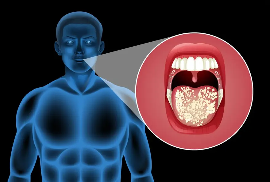
A yellow tongue, characterised by a yellowish coating on the tongue's surface, is a relatively common condition that can cause concern. While poor oral hygiene is often the culprit, leading to a buildup of bacteria, dead cells, and debris, a yellow tongue may occasionally signal an underlying health issue. In this article, we'll delve into yellow tongue causes and symptoms, how it's diagnosed, and the treatment options available. Our goal is to equip you with the knowledge to identify a yellow tongue, understand its implications, and take appropriate steps to restore your tongue to a healthy pink. Whether you're dealing with yellow tongue due to lifestyle factors like smoking or poor brushing habits, or have concerns about a potential medical condition, this guide will provide the clarity and direction you need. Why is our tongue yellow? A yellow tongue occurs when there is a buildup of dead skin cells, bacteria, yeast, tobacco stains, or other debris on the tongue's surface. Poor oral hygiene is a primary culprit, allowing this yellowish film to accumulate over time. Certain foods and drinks, like coffee and tea, contain staining particles that can also contribute to discolouration. In some cases, a dry mouth can exacerbate the issue by reducing saliva flow, which normally helps cleanse the tongue. Less commonly, an overgrowth of papillae or an underlying medical condition may cause the tongue to take on a yellowish hue. What does a yellowish tongue mean? A yellow tongue is often an indication of poor oral hygiene habits or exposure to staining substances. However, it can occasionally be a warning sign of an underlying health problem. Conditions like acid reflux, bacterial or yeast infections, and in rare cases, jaundice related to liver issues, may manifest with tongue discolouration. Who does yellow tongue affect? Yellow tongue causes can impact people of all ages, but is more common in individuals with suboptimal oral hygiene routines, tobacco users, and those with certain health conditions or taking specific medications. What causes yellow tongue? Several factors can contribute to the development of yellow tongue causes, ranging from lifestyle habits to medical issues: Poor oral hygiene: Failing to brush and floss regularly allows bacteria, fungi, and dead cells to accumulate on the tongue's surface, leading to a yellowish buildup. Tobacco use: Smoking or chewing tobacco can stain the tongue yellow due to the tar and nicotine content. Certain foods and drinks: Consuming coffee, tea, red wine, and deeply colored berries can leave staining particles on the tongue. Dry mouth: Conditions that reduce saliva production, such as dehydration, mouth breathing, or medications with dry mouth as a side effect, can allow debris to collect on the tongue more easily. Medications: Some antibiotics, antipsychotics, and antihypertensives may cause tongue discolouration as a side effect. Oral thrush: This yeast infection, caused by an overgrowth of Candida albicans, can create white or yellowish patches on the tongue. Black hairy tongue: A harmless condition where the papillae become elongated and trap bacteria and stains, sometimes appearing yellowish before turning dark. Geographic tongue: This benign condition causes patches of papillae to disappear, leaving irregular red areas that may be bordered by a yellow or white coating. Gastric reflux: Acid backing up from the stomach can irritate the tongue and contribute to discolouration. Jaundice: In rare cases, a yellowing of the tongue might indicate a buildup of bilirubin related to liver disease. It's important to note that a yellow tongue diagnosis is usually harmless and resolves with proper oral care. However, if the discolouration persists, is accompanied by pain or other unusual symptoms, or you suspect an underlying condition, it's best to consult a healthcare professional for an accurate diagnosis and appropriate treatment plan. What are the symptoms of yellow tongue? Among yellow tongue symptoms, the primary sign is a noticeable yellowish coating or film on the tongue's surface. This discolouration can range from a light, barely visible yellow to a thicker, more pronounced layer. Some people may also experience: Bad breath or an unpleasant taste in the mouth A burning or tingling sensation on the tongue Dry mouth or excessive thirst Sore throat or difficulty swallowing Papillae that appear longer or larger than normal Cracks or grooves on the tongue's surface In rare cases, a yellow tongue may be accompanied by more severe symptoms like: Fever or chills Persistent pain or tenderness in the tongue Difficulty speaking or moving the tongue Yellowing of the skin or eyes (jaundice) If you experience any of these additional yellow tongue symptoms, it's crucial to seek medical attention promptly, as they could indicate a more serious underlying condition requiring treatment. How is yellow tongue diagnosed? A yellow tongue diagnosis typically involves a visual examination by a healthcare provider. They will assess the tongue's appearance, texture, and any accompanying symptoms. In some cases, a detailed medical history and additional tests like blood work or oral swabs may be necessary to rule out underlying conditions. How do you get rid of a yellow tongue? Yellow tongue treatment depends on the underlying cause but often includes simple home remedies and lifestyle changes: Improve oral hygiene: Brush your teeth twice daily, floss regularly, and gently brush your tongue or use a tongue scraper to remove buildup. Quit smoking: Eliminating tobacco use can prevent further staining and promote overall oral health. Stay hydrated: Drinking plenty of water can help stimulate saliva production and prevent dry mouth. Limit staining foods and drinks: Reduce your intake of coffee, tea, and deeply pigmented foods that may contribute to discolouration. Manage underlying conditions: If your yellow tongue symptoms are related to acid reflux, thrush, or another medical issue, treating the root cause can help resolve the discoloration. In most cases, a combination of good oral hygiene practices and lifestyle modifications can effectively treat a yellow tongue. If the discolouration persists or you have concerns, consult your dentist or doctor for further guidance. Can we prevent yellow tongue? Preventing a yellow tongue involves adopting healthy oral hygiene habits and making some lifestyle adjustments: Brush and floss regularly: Maintain a consistent oral care routine, brushing your teeth twice a day and flossing daily to remove plaque and bacteria. Use a tongue scraper: Gently scraping your tongue once or twice a day can help remove the buildup of debris and prevent discolouration. Quit smoking: Avoiding tobacco products can significantly reduce your risk of developing a yellow tongue and other oral health issues. Limit staining foods and drinks: Reduce your consumption of coffee, tea, red wine, and deeply pigmented foods that can contribute to tongue discolouration. Stay hydrated: Drinking water throughout the day can help stimulate saliva flow, which naturally cleanses the tongue and prevents dry mouth. Manage underlying conditions: If you have acid reflux, diabetes, or another chronic condition that may contribute to yellow tongue, work with your healthcare provider to keep it well-controlled. Schedule regular dental check-ups: Visit your dentist for professional cleanings and exams every six months to maintain optimal oral health and catch any potential issues early. What's the outlook for people with yellow tongue? For most people, a yellow tongue diagnosis is a harmless and temporary condition that resolves with proper oral hygiene and lifestyle changes. Once the underlying cause is addressed, the tongue typically returns to its normal color within a few weeks. However, if the discolouration persists despite self-care measures, or if you experience pain, difficulty swallowing, or other concerning symptoms, it's essential to consult a healthcare professional. In rare cases, a yellow tongue symptoms may indicate a more serious underlying condition that requires prompt medical attention and treatment. When to see a Doctor While a yellow tongue is usually benign and can be managed at home, there are certain situations where seeking medical advice is necessary: Persistent discolouration: If your yellow tongue does not improve after a few weeks of proper oral hygiene and lifestyle changes, consult your dentist or doctor. Pain or discomfort: If your tongue becomes painful, tender, or swollen, or if you experience difficulty swallowing or speaking, seek medical attention. Other symptoms: If your yellow tongue is accompanied by fever, chills, or other concerning symptoms, consult a healthcare professional promptly. Jaundice: If you notice a yellowing of your skin or eyes along with your yellow tongue, seek immediate medical care, as this may indicate a serious liver issue. Conclusion In conclusion, a yellow tongue is a common condition that can cause concern but is usually harmless and easily treatable. By understanding the various causes, from poor oral hygiene to underlying health issues, you can take proactive steps to prevent and manage tongue discoloration. At Metropolis Healthcare, we understand the importance of oral health in overall well-being. Our team of skilled pathologists and technicians offer a wide range of diagnostic tests, including those related to underlying conditions that may cause yellow tongue. With our convenient at-home sample collection service, you can have your blood or other samples taken in the comfort of your own home, and access your test results online via email or our user-friendly Metropolis TruHealth app.
Blood In Semen (Hematospermia): Causes & Treatment
Noticing blood in your semen can be an alarming experience that leaves you with many questions and concerns. This condition, medically known as hematospermia or blood in semen, is often benign but can sometimes indicate an underlying health issue. While the sight of blood in semen may cause distress, understanding the potential causes can help ease anxiety. Blood in semen causes may include factors such as infections, inflammation, or minor injuries to the reproductive tract. In some cases, medical procedures like biopsies can also lead to this condition. Diagnosing hematospermia typically involves a semen analysis, which helps identify signs of infection, inflammation, or other abnormalities. Fortunately, blood in semen treatment options are often straightforward, especially when an infection or minor injury is the cause. Antibiotics, anti-inflammatory medications, or lifestyle adjustments may be recommended depending on the diagnosis. However, if blood in your semen persists or is accompanied by pain, swelling, or urinary issues, seeking medical advice is crucial. By understanding more about this condition and exploring available treatments, you can take proactive steps toward your well-being and peace of mind. What is blood in semen? Hematospermia, or blood in the semen, occurs when semen appears pink, red, or brown due to the presence of blood. This can range from faint streaks to more noticeable discolouration. While blood in the semen can be alarming, it's often harmless and resolves without treatment. However, in some cases, it may indicate an underlying issue within the male reproductive system. If the condition persists or is accompanied by other symptoms, seeking medical advice is recommended to identify potential causes and ensure proper care. Should we worry about blood in our semen? In most cases, hematospermia is not a cause for major concern and resolves without treatment. However, if you experience persistent or recurrent blood in your semen, it's important to consult a healthcare provider. This is especially true for men over 40 or those with risk factors for prostate issues, as it may indicate an underlying condition that requires medical attention. Is blood in semen a common condition? While not extremely common, hematospermia does occur in men of all ages. A study published in the Journal of Sexual Medicine found that about 1 in 5,000 men seek medical attention for blood in their semen each year. Infections of the prostate or urethra are among the most frequent causes. Is seeing blood in our semen normal? Occasional episodes of hematospermia, particularly after prolonged sexual abstinence or vigorous sexual activity, may not be abnormal. However, if you notice blood in your semen regularly or if it's accompanied by pain, discomfort, or other concerning symptoms, it's best to consult a doctor for proper evaluation and guidance. Why would I have blood in my semen? There are several potential causes of hematospermia, ranging from minor concerns to more serious conditions that require medical evaluation. Understanding these causes can help determine whether medical attention is necessary. Infections are one of the most common blood in semen causes. Conditions like prostatitis (inflammation of the prostate) and urethritis (inflammation of the urethra) frequently lead to blood in semen. These infections may be triggered by bacteria, viruses, or sexually transmitted infections (STIs) such as chlamydia or gonorrhoea. Infections can also cause discomfort, pain during ejaculation, or urinary symptoms. Treating the infection typically resolves the issue. Trauma is another frequent cause. Injuries resulting from vigorous sexual activity, masturbation, or recent medical procedures like prostate biopsies, vasectomies, or catheter insertions can damage blood vessels and cause bleeding. This type of hematospermia is often temporary and clears up within a few weeks. Tumours are a less common but more serious cause. Prostate, testicular, or urethral cancers can occasionally present with blood in semen. This is more likely in older men or those with additional risk factors such as unexplained weight loss, pelvic pain, or urinary issues. Early diagnosis through regular health screenings can improve outcomes. Structural abnormalities in the seminal vesicles, ejaculatory ducts, or reproductive organs can also cause bleeding. These may include cysts, blockages, or congenital malformations that irritate surrounding tissues. Vascular issues are another possible cause. Fragile or abnormal blood vessels in the prostate or seminal vesicles may rupture, especially in men with conditions like hypertension or those on blood-thinning medications. Other less common causes include systemic conditions such as bleeding disorders, liver disease, or severe hypertension. Certain medications that affect blood clotting, like anticoagulants, may also increase the risk of hematospermia. Can a hernia cause blood in my semen? Hernias, which involve the protrusion of tissue through a weak spot in the abdominal wall, do not directly cause hematospermia. Blood in semen typically originates from issues within the reproductive or urinary tract. What does brown blood in semen mean? Brown blood in semen suggests that the bleeding occurred some time ago, allowing the blood to oxidise and darken in colour. This may indicate a chronic or low-grade bleeding issue within the reproductive tract. It's important to bring this up with your healthcare provider for a proper evaluation. How is blood in semen diagnosed? Diagnosing hematospermia causes typically involves a combination of the following: Medical history: Your doctor will ask about your symptoms, sexual history, and any recent injuries or medical procedures. Physical examination: A thorough examination of the genitals, prostate gland, and abdomen can help identify any abnormalities or signs of infection. Semen analysis: A laboratory evaluation of your semen can detect the presence of blood, assess sperm health, and check for signs of infection. Urine tests: Analysing a urine sample can help diagnose urinary tract infections or STIs that may be causing blood in the semen. Imaging tests: In some cases, your doctor may recommend an ultrasound, CT scan, or MRI to visualise the reproductive organs and check for structural issues or tumours. Blood tests: Evaluating blood samples can help rule out systemic conditions like bleeding disorders or assess prostate health through tests like the prostate-specific antigen (PSA) test. How do you treat bloody semen? Blood in semen treatment depends on the underlying cause identified through diagnostic tests. Some common approaches include: Antibiotics: If an infection is responsible for the hematospermia, antibiotics will be prescribed to clear the infection and alleviate symptoms. Watchful waiting: In many cases, blood in the semen resolves on its own without specific treatment. Your doctor may recommend monitoring the situation and practicing abstinence until the bleeding subsides. Surgical intervention: If a structural abnormality or tumour is causing the bleeding, surgical correction or removal may be necessary. Medication adjustments: If a medication is contributing to hematospermia, your doctor may recommend adjusting the dosage or switching to an alternative drug. Should we stop masturbating if we have blood in our semen? While excessive or vigorous masturbation can potentially irritate the reproductive tract and cause temporary hematospermia, completely stopping masturbation is not necessary unless advised by your healthcare provider. The key is to identify the underlying cause through a proper medical evaluation and follow your doctor's recommendations. What are the possible complications or risks of not treating hematospermia? In most cases, untreated hematospermia does not lead to serious complications. However, if an underlying infection or cancer is causing the blood in semen and is left unaddressed, it can potentially lead to: Spread of the infection to other parts of the body Damage to the reproductive organs Infertility Progression of cancerous growths When to see a Doctor It's important to consult a doctor if you experience any of the following: Persistent or recurrent episodes of hematospermia Blood in the semen accompanied by pain, fever, chills, or other concerning symptoms Difficulty urinating or painful ejaculation Swelling or lumps in the testicles or prostate area Unexplained weight loss or fatigue Men over 40 or those with risk factors for prostate issues should be especially proactive about seeking medical evaluation and subsequent blood in semen treatment. Conclusion While discovering blood in your semen can be unnerving, remember that most cases of hematospermia are benign and treatable. By understanding the potential causes, seeking timely medical advice, and following through with recommended diagnostic tests and blood in semen treatment, you can take charge of your reproductive health. If you're experiencing concerning symptoms or have questions about semen analysis and other diagnostic procedures, consider reaching out to Metropolis Healthcare. With a network of advanced diagnostic labs across India and a commitment to delivering reliable results and personalised care, Metropolis Healthcare can be your trusted partner in navigating this sensitive issue and prioritising your well-being.
Vaginal Odor: Types, Causes, Diagnosis & Treatment
Vaginal odor is a common concern for many women, yet it’s often misunderstood due to stigma. While a mild scent is normal, certain odors may signal underlying health issues. Understanding the causes, types, and treatment of vaginal odor can help you manage your intimate health with confidence. Factors such as hygiene, diet, and hormonal changes can influence odor, but persistent or unusual smells may require medical attention. This blog explores common causes, how to identify concerning odors, and when to seek professional advice. By staying informed and proactive, you can maintain optimal reproductive health and overall well-being. Remember, your body’s signals are important—knowing what’s normal and what’s not empowers you to take control of your health. What is abnormal vaginal odor? Abnormal vaginal odor refers to a strong, unpleasant, or unusual scent that deviates from your natural smell. While a mild, musky odor is normal, foul, fishy, or rotten smells may indicate an imbalance or infection. Common vaginal odor causes include bacterial vaginosis, yeast infections, or poor hygiene. Hormonal changes, certain foods, and medications can also affect vaginal odor. If the odor is persistent, accompanied by itching, discharge, or discomfort, it’s important to seek medical advice for proper diagnosis and treatment. What causes vaginal odor? Vaginal odor causes can result from various factors, including bacterial vaginosis (BV), trichomoniasis, and yeast infections like vulvovaginal candidiasis. Poor hygiene practices, hormonal changes during menstrual cycles or menopause, and certain foods or medications can also contribute. Identifying the root cause is key to finding the right treatment. While some triggers are temporary and manageable, persistent or strong odors may signal an infection or imbalance. Understanding these causes empowers you to take proactive steps toward maintaining vaginal health. Normal vaginal odors It's important to understand that some vaginal odor types are entirely normal and can vary between individuals. Typically described as musky or slightly sweet, normal vaginal odors may change throughout your menstrual cycle due to hormonal fluctuations. Factors such as diet, hygiene habits, and clothing choices can also influence these natural scents. For instance, foods like garlic, onions, or asparagus may temporarily alter vaginal odor. Likewise, wearing tight, non-breathable fabrics can trap moisture, intensifying the scent. However, these odors are usually mild and should not be overpowering or unpleasant. To determine if your vaginal odor is within the normal range, consider the following: Is the odor subtle and not unusually strong? Does it change slightly throughout your menstrual cycle? Is it free from other symptoms such as itching, burning, or unusual discharge? If you answered yes to these questions, your vaginal odor is likely normal. It's also helpful to remember that post-exercise sweat or sexual activity can temporarily alter vaginal scent without indicating a problem. However, if you notice a persistent, strong, or foul odor—especially when paired with discomfort or unusual discharge—it’s advisable to consult a healthcare provider for assessment and guidance. Understanding what’s normal can help you feel more confident in managing your intimate health. Abnormal vaginal odors Abnormal vaginal odors can vary in scent, and identifying the vaginal odor type can help uncover potential health concerns. While some changes are temporary and harmless, persistent or strong odors may require medical attention. Fishy odor: A strong, fishy smell is commonly linked to bacterial vaginosis (BV), caused by an overgrowth of anaerobic bacteria. This odor may intensify after sexual intercourse or during menstruation. BV may also cause thin, greyish-white discharge. Foul or rotten odor: A foul, rotten smell can indicate trichomoniasis, a sexually transmitted infection (STI). This condition often presents with a greenish, frothy discharge and vaginal discomfort. Prompt treatment is essential to prevent complications. Yeasty or beer-like odor: A sweet, beer-like scent may suggest vulvovaginal candidiasis (yeast infection), triggered by an overgrowth of fungus. Yeast infections are commonly accompanied by itching, irritation, and thick, white discharge resembling cottage cheese. Strong, musky odor: While mild musky scents are normal, an intense or persistent musky odor—especially one that lingers despite proper hygiene—may indicate an underlying issue. If you notice abnormal vaginal odors, especially with symptoms like itching, burning, or unusual discharge, consult a healthcare provider. Early vaginal odor diagnosis and treatment are crucial to maintaining vaginal health and preventing further complications. What causes vaginal odor during pregnancy? Vaginal odor during pregnancy can be influenced by the hormonal changes that occur during this period. This is usually normal and not a cause for concern. However, pregnant women are more susceptible to certain infections, such as bacterial vaginosis and yeast infections, which can cause abnormal vaginal odors. Increased estrogen levels and increased blood flow to the vaginal area can alter the vaginal pH, making it more susceptible to bacterial overgrowth and infections like BV. Additionally, pregnancy can cause changes in vaginal discharge, which may contribute to odor. It's important for pregnant women to maintain good hygiene practices, wear breathable clothing, and avoid irritants to help manage vaginal odor. If you experience persistent, strong odors accompanied by other symptoms during pregnancy, consult your healthcare provider to rule out any infections or complications that may impact your health or the health of your baby. Tips for reducing unpleasant vaginal odors To minimise unpleasant vaginal odor: Practice good hygiene by gently cleansing the external genital area daily with mild soap and water Wear breathable, cotton underwear and avoid tight-fitting clothing Change out of wet or sweaty clothing promptly Wipe from front to back after using the restroom to avoid introducing harmful bacteria into the vagina How is abnormal vaginal odor diagnosed? Vaginal odor diagnosis typically involves a pelvic exam and a review of your medical history. Your healthcare provider may also collect samples of vaginal discharge for laboratory testing, such as: Wet mount: A sample of vaginal discharge is examined under a microscope to identify the presence of bacteria, yeast, or parasites. Vaginal pH test: Measuring the pH level of the vagina can help determine if there is an imbalance in the vaginal flora. Vaginal cultures: In some cases, a culture may be necessary to identify specific bacteria or fungi causing the odor. STI testing: If a sexually transmitted infection is suspected, your healthcare provider may recommend tests for gonorrhoea, chlamydia, or trichomoniasis. Based on the results of these diagnostic tests, your healthcare provider can determine the underlying cause of the abnormal odor and recommend an appropriate vaginal odor treatment plan. How is vaginal odor treated? Vaginal odor treatment depends on the specific cause identified through diagnosis. Common treatment approaches include: Antibiotics: If bacterial vaginosis or trichomoniasis is diagnosed, your healthcare provider may prescribe oral or vaginal antibiotics to eliminate the infection. Antifungal medications: Yeast infections can be treated with over-the-counter or prescription antifungal creams, suppositories, or oral medications. Hygiene modifications: In some cases, making adjustments to hygiene practices, such as avoiding scented products or changing underwear more frequently, can help resolve vaginal odor. Treating underlying conditions: If the odor is a result of an underlying medical condition, such as diabetes or a hormonal imbalance, treating the root cause can help alleviate the symptom. It's crucial to complete the full course of any prescribed treatment and follow your healthcare provider's recommendations to ensure the effectiveness of vaginal odor treatment. How can vaginal odor be prevented? Preventing vaginal odor involves a combination of good hygiene practices and lifestyle habits. Some key prevention strategies include: Maintaining cleanliness: Gently wash the external vaginal area daily with mild, unscented soap and warm water. Avoid douching or using scented products, as these can disrupt the natural vaginal flora. Wearing breathable clothing: Choose cotton underwear and avoid tight-fitting clothing that can trap moisture and promote bacterial growth. Practising safe sex: Using condoms during sexual activity can help prevent the introduction of bacteria or sexually transmitted infections that can cause odor. Staying hydrated: Drinking plenty of water helps maintain the body's natural balance and can help prevent odor-causing bacteria from thriving. Wiping from front to back: After using the restroom, always wipe from front to back to avoid introducing bacteria from the anus to the vaginal area. By incorporating these preventive measures into your daily routine, you can help maintain a healthy vaginal environment and reduce the risk of developing abnormal vaginal odor. When to see a doctor If you experience persistent, strong vaginal odors accompanied by other symptoms, it's important to consult a healthcare provider promptly. Some signs that warrant medical attention include: Intense, foul-smelling odor that does not resolve with hygiene measures Greenish, greyish, or bloody vaginal discharge Itching, burning, or irritation in the vaginal area Pain during urination or sexual intercourse These symptoms can indicate an underlying infection or condition that requires treatment. Seeking early intervention can help prevent complications and promote faster recovery. Additionally, if you are pregnant and experience any concerning changes in vaginal odor or discharge, inform your healthcare provider immediately. Certain infections during pregnancy can pose risks to both the mother and the developing foetus, making prompt diagnosis and treatment essential. Remember, your healthcare provider is there to help you navigate any concerns about your vaginal health. Don't hesitate to ask questions or voice your concerns during your appointment. By working together, you can develop an effective plan to manage and prevent abnormal vaginal odor. Conclusion Vaginal odor is a common concern that can cause significant distress and embarrassment for many women. By understanding the different vaginal odor types, their causes, and the available diagnostic and treatment options, you can take control of your reproductive health. At Metropolis Healthcare, we understand the importance of accurate diagnosis in addressing vaginal health concerns. Our team of skilled technicians provides reliable pathology testing services to help identify the underlying causes of abnormal vaginal odor. With our convenient at-home sample collection and online report delivery, you can prioritise your health with ease. FAQs How do I stop smelling down there? To reduce vaginal odor, practice good hygiene by gently cleansing the external vaginal area daily with mild soap and warm water. Avoid using scented products or douching, as these can disrupt the natural vaginal flora. Wear breathable, cotton underwear and change out of wet or sweaty clothing promptly. Also, wiping from front to back after using the restroom helps prevent the condition. If odors persist despite these measures, consult a healthcare provider for further evaluation. Why do I have a strong odor down there? A strong vaginal odor can be caused by bacterial vaginosis, trichomoniasis, or yeast infections. Hormonal changes, poor hygiene, certain foods, or medications may also play a role. While some scent changes are normal, a persistent, strong odor—especially with symptoms like itching, burning, or unusual discharge—may indicate an infection or imbalance. Maintaining proper hygiene, wearing breathable fabrics, and avoiding harsh soaps can help. If the odor persists, consult a healthcare provider for diagnosis and appropriate treatment. What does BV smell like? Bacterial vaginosis (BV) is commonly linked to a strong, fishy odor caused by an overgrowth of anaerobic bacteria in the vagina. This odor may become more noticeable after sexual intercourse or during menstruation. BV can also cause thin, greyish-white discharge. If you suspect BV, it's important to consult a healthcare provider for proper evaluation and treatment. Timely care can help restore vaginal balance and prevent potential complications, ensuring your intimate health remains well-managed.
Retrograde Ejaculation: Causes, Symptoms, and Treatment Options
Retrograde ejaculation is a condition that affects male sexual health and can lead to fertility issues. If you or your partner are experiencing dry orgasms or cloudy urine after ejaculation, it's essential to understand the underlying causes, symptoms, and available treatment options. This article will provide you with a comprehensive overview of retrograde ejaculation, helping you make informed decisions about your sexual health and fertility. What is retrograde ejaculation? Retrograde ejaculation occurs when semen, which normally exits through the penis during orgasm, instead flows backwards into the bladder. This happens due to a failure of the bladder neck muscles to contract and close off the bladder during ejaculation. As a result, semen mixes with urine in the bladder, leading to little or no semen being released during orgasm. Who does retrograde ejaculation affect? Retrograde ejaculation can affect men of any age, but it is more common in those with certain medical conditions or who have undergone specific treatments. Some factors that increase the risk of developing retrograde ejaculation include: Diabetes Prostate surgery, such as transurethral resection of the prostate (TURP) Spinal cord injuries Multiple sclerosis Certain medications, such as alpha-blockers or antidepressants How common is retrograde ejaculation? Retrograde ejaculation is relatively uncommon, affecting about 0.3% to 2% of men. However, its prevalence can vary based on the cause. For instance, up to 75% of men who undergo TURP surgery may experience retrograde ejaculation afterward. While rare in the general population, this condition is more frequent in individuals with specific medical procedures or conditions. Understanding the risk factors can help with early retrograde ejaculation diagnosis and management. What are the signs and symptoms of retrograde ejaculation? The primary retrograde ejaculation symptoms and signs include: Little or no semen during ejaculation (dry orgasm) Cloudy urine after sexual activity, due to the presence of semen in the bladder Inability to conceive, as semen does not exit the penis during intercourse It's important to note that men with retrograde ejaculation still experience normal orgasms and sexual pleasure, despite the absence of semen. What causes retrograde ejaculation? Several factors can contribute to the development of retrograde ejaculation causes, including: Diabetes: Nerve damage caused by poorly controlled diabetes can affect the muscles controlling ejaculation. Medications: Certain drugs, such as alpha-blockers for treating high blood pressure and prostate enlargement, antidepressants, and antipsychotics, can relax the bladder neck muscles and lead to retrograde ejaculation. Surgery: Procedures involving the prostate, bladder, or urethra can damage the nerves and muscles responsible for normal ejaculation. Nerve damage: Conditions like multiple sclerosis, Parkinson's disease, and spinal cord injuries can disrupt nerve signals involved in ejaculation. How is retrograde ejaculation diagnosed? If you suspect you have the condition, consult a healthcare provider for an accurate retrograde ejaculation diagnosis. The diagnostic process typically involves: Medical history review: Your doctor will ask about your symptoms, sexual history, and any medications or surgeries you've had. Physical examination: A thorough examination of the genitals and prostate can help identify any structural issues or abnormalities. Post-ejaculation urine analysis: A sample of your urine collected shortly after ejaculation will be examined for the presence of sperm, confirming retrograde ejaculation. What are some complications or side effects related to drug treatment of retrograde ejaculation? Medications like pseudoephedrine and imipramine can help with retrograde ejaculation treatment by stimulating the bladder neck muscles to contract during ejaculation. However, these drugs may cause side effects such as: Increased heart rate and blood pressure Anxiety and restlessness Difficulty sleeping Dry mouth and nausea Are there exercises that help with retrograde ejaculation? While there are no specific exercises proven to be beneficial for retrograde ejaculation treatment, maintaining overall pelvic floor health through regular Kegel exercises may be helpful. These exercises involve contracting and relaxing the muscles used to control urination and can help improve bladder control and sexual function. How can we prevent retrograde ejaculation? Preventing retrograde ejaculation depends on the underlying cause. Some strategies include: Managing diabetes through proper diet, exercise, and medication to prevent nerve damage Discussing alternative medications with your doctor if you suspect your current drugs are causing retrograde ejaculation Considering nerve-sparing surgical techniques, when possible, to minimize damage during prostate or bladder procedures Maintaining a healthy lifestyle to promote overall sexual and urological health What is the outlook for retrograde ejaculation? The prognosis for retrograde ejaculation varies depending on the cause and individual circumstances. In some cases, such as when the condition is caused by medication, stopping or changing the drug may resolve the issue. However, if nerve damage or surgical complications are responsible, retrograde ejaculation may be permanent. When to see a doctor? If you experience retrograde ejaculation symptoms, particularly if you and your partner are trying to conceive, it's important to consult a healthcare provider. They can help diagnose the underlying cause, discuss potential treatment options, and provide guidance on managing the condition and optimising your sexual health and fertility. Conclusion Retrograde ejaculation is a manageable condition that requires proper diagnosis and treatment to address its impact on sexual health and fertility. If you suspect you have retrograde ejaculation or are experiencing any concerning symptoms, don't hesitate to seek medical advice. Metropolis Healthcare, a leading chain of diagnostic labs across India, offers comprehensive pathology testing and health check-up services to help you prioritise your health. With a team of qualified blood collection technicians and advanced diagnostic labs, Metropolis Healthcare is committed to delivering reliable results and personalised care. FAQs What does retrograde ejaculation feel like? Men with retrograde ejaculation still experience normal orgasms and sexual pleasure, but they may notice little to no semen during ejaculation. This is often described as a "dry orgasm." Does retrograde ejaculation go away? The likelihood of retrograde ejaculation resolving depends on the underlying cause. If the condition is due to medication, stopping or changing the drug may reverse the issue. However, if nerve damage or surgical complications are responsible, retrograde ejaculation may be permanent. How common is it after TURP surgery? Retrograde ejaculation is a common side effect of transurethral resection of the prostate (TURP) surgery, with studies showing that up to 75% of men experience the condition post-operatively. Are there benefits to retrograde ejaculation? While there are no direct benefits to retrograde ejaculation, it's important to note that the condition does not affect sexual pleasure or performance. Men with retrograde ejaculation can still have satisfying sexual experiences, despite the absence of semen during ejaculation.
What is Parkinsonism? Types, Symptoms, and Treatment Approaches
What is Parkinsonism? Parkinsonism refers to a group of neurological disorders that cause movement symptoms similar to those seen in Parkinson's disease. However, while Parkinson's is a specific disease, parkinsonism is an umbrella term for conditions that lead to Parkinson's-like symptoms. Parkinsonism can result from various causes, such as side effects of certain medications, brain injuries, strokes, or neurodegenerative disorders other than Parkinson's disease. Identifying the underlying cause is crucial for determining the appropriate treatment approach. If you or a loved one are experiencing symptoms of Parkinsonism, or Parkinson's disease, it's essential to consult a neurologist for an accurate diagnosis. Parkinsonism vs. Parkinson's Disease: Key Differences While Parkinsonism and Parkinson's disease share similar symptoms, there are important differences between the two conditions: Features Parkinson's Disease Parkinsonism Primary Cause Degeneration of dopamine-producing brain cells Various conditions (medications, brain injuries, etc.) Symptoms Progressive tremors, stiffness, slow movements Similar symptoms plus others based on underlying cause Treatment Response Responds well to levodopa medication Often less responsive to levodopa Progression Gradually worsening over time Varies depending on the specific cause Unlike Parkinson's disease, which primarily involves the death of dopamine-producing cells in a part of the brain called the substantia nigra, Parkinsonism can have diverse causes. These may include: Side effects of certain medications, especially antipsychotics and anti-nausea drugs Brain injuries or tumours Strokes affecting the brain's movement control centres Neurodegenerative disorders like multiple system atrophy or progressive supranuclear palsy Types of Parkinsonism Parkinsonism can be classified into several types based on the underlying cause: Drug-induced Parkinsonism: Certain medications, particularly antipsychotics used to treat psychiatric disorders and anti-nausea drugs like metoclopramide, can cause parkinsonian symptoms as a side effect. These symptoms usually resolve once the offending medication is discontinued. Vascular Parkinsonism: This type results from one or more small strokes that damage the brain's movement control centers. The symptoms may appear more suddenly compared to the gradual onset of Parkinson's disease. Post-traumatic Parkinsonism: Head injuries, especially those involving damage to the basal ganglia or midbrain regions, can lead to Parkinson's symptoms. Boxers and football players are at an increased risk due to repeated concussions. Toxin-induced Parkinsonism: Exposure to certain toxins, such as carbon monoxide, cyanide, and manganese, can cause damage to the brain's movement control circuits, resulting in Parkinsonism. Multiple system atrophy (MSA): This neurodegenerative disorder affects multiple brain areas, leading to parkinsonism along with impaired autonomic functions like blood pressure control and bladder function. Progressive supranuclear palsy (PSP): PSP is characterised by Parkinsonism, eye movement abnormalities, and cognitive decline. Falls are a common early symptom in this condition. Identifying the specific type of Parkinsonism or types of Parkinson's disease is crucial for determining the most appropriate Parkinson's disease treatment approach. Symptoms of Parkinsonism The symptoms of Parkinsonism can vary based on the underlying cause, but common signs include: Tremors: Involuntary shaking, usually starting in the hands, fingers, or legs. These tremors are most noticeable at rest and may lessen with movement. Rigidity: Muscle stiffness that limits flexibility and can cause discomfort. Muscles may feel tight and contracted. Bradykinesia: Slowness of movement, making everyday tasks like dressing and eating more challenging. Postural Instability: Poor balance and coordination, leading to a stooped posture and increased fall risk. Shuffling Gait: Walking with short, shuffling steps and reduced arm swing. Some early symptoms of Parkinson's disease may also include: Reduced facial expressions (facial masking) Soft or hoarse voice Difficulty swallowing Constipation Sleep disturbances Cognitive changes, such as slowed thinking and attention difficulties It's important to note that not everyone with parkinsonism will experience all these symptoms, and the severity can vary from person to person. If you notice any of these Parkinson's symptoms in yourself or a loved one, it's essential to consult a neurologist promptly for a thorough evaluation. Causes and Risk Factors Parkinsonism is a complex neurological syndrome that affects movement and balance. While Parkinson's disease is the most common cause of Parkinsonism, several other conditions can lead to similar symptoms. Understanding the underlying Parkinson's disease causes and other Parkinsonian disorders is crucial for early detection and effective management. Idiopathic Parkinson's disease, which accounts for the majority of Parkinsonism cases, has no known specific cause. However, researchers believe that a combination of genetic and environmental factors may contribute to its development. Vascular Parkinsonism results from reduced blood flow to the brain, often due to small strokes or chronic cerebrovascular disease. Drug-induced Parkinsonism can occur as a side effect of certain medications, such as antipsychotics and calcium channel blockers. Parkinson-plus syndromes, like multiple system atrophy and progressive supranuclear palsy, involve additional neurological symptoms beyond typical Parkinsonian features. Age remains the most significant risk factor for Parkinsonism, with incidence increasing dramatically after age 60. Other potential risk factors include family history of Parkinson's disease or related disorders, exposure to environmental toxins like pesticides and heavy metals, or head trauma or brain injury. Diagnosis of Parkinsonism Diagnosing Parkinsonism requires a thorough clinical evaluation by a neurologist or movement disorder specialist. There is no single definitive Parkinson's test, so diagnosis relies on identifying characteristic symptoms and ruling out other potential causes. The diagnostic process typically involves: Detailed medical history to assess symptom onset, progression, and risk factors Physical and neurological examination to evaluate Parkinsons symptoms like tremor, rigidity, and bradykinesia Cognitive and psychiatric assessment to detect non-motor symptoms Brain imaging tests (MRI, PET, DaTscan) to visualize brain structure and function In some cases, a levodopa challenge may be performed to assess responsiveness to dopamine replacement therapy, which can help differentiate idiopathic Parkinson's disease from other Parkinsonian syndromes. However, confirming a specific diagnosis often requires monitoring symptom progression over time. If you notice any early symptoms of Parkinson's disease, such as tremor, stiffness, or changes in gait, it's essential to consult a healthcare provider for a proper evaluation. Treatment Options for Parkinsonism While there is no cure for Parkinsonism, various Parkinson's treatment approaches can help manage symptoms and improve quality of life. The primary goals of treatment are to: Alleviate motor symptoms like tremor, rigidity, and slow movement Address non-motor symptoms such as depression, anxiety, and sleep disturbances Maintain independence and functional ability for as long as possible Medications Medication is the cornerstone of Parkinson's disease treatment. The most effective drug is Levodopa, often combined with Carbidopa. Other medications include: Dopamine Agonists (Ropinirole, Pramipexole) MAO-B Inhibitors (Selegiline, Rasagiline) COMT Inhibitors (Entacapone, Tolcapone) Anticholinergics (Trihexyphenidyl) Surgical Treatment For advanced cases, Deep Brain Stimulation (DBS) may be recommended. This procedure involves implanting electrodes in the brain to regulate abnormal activity. Complementary Therapies Physical Therapy: Enhances mobility, balance, and flexibility Speech Therapy: Helps with communication and swallowing difficulties Occupational Therapy: Supports daily activities and home safety A personalised Parkinson's disease treatment plan will be developed based on symptoms, disease stage, and individual needs. Living with Parkinsonism: Management and Coping Strategies In addition to medical treatment, making certain lifestyle adjustments can go a long way in managing Parkinson's symptoms and coping with the challenges of living with a chronic neurological condition. Consider implementing the following tips: Stay active with regular exercise tailored to your abilities, such as walking, swimming, or cycling. Physical activity can help improve mobility, balance, and mood. Eat a balanced diet rich in fiber, antioxidants, and omega-3 fatty acids. Adequate nutrition supports brain health and overall well-being. Practice stress-reduction techniques like deep breathing, meditation, or gentle yoga to relax muscles and calm the mind. Prioritise sleep hygiene by establishing a consistent sleep schedule, creating a comfortable sleep environment, and avoiding stimulants late in the day. Install handrails, grab bars, and non-slip mats in the bathroom and shower. Use utensils with large handles or weighted utensils for easier gripping. Consider a walker or wheelchair for safe mobility if needed. Remember, you don't have to face Parkinsonism alone. Seek support from family, friends, and local Parkinson's organisations. Conclusion Parkinsonism encompasses a spectrum of neurological disorders that share common features of tremor, stiffness, and slow movement. While Parkinson's disease is the most prevalent cause, various other conditions can lead to Parkinsonian symptoms. If you notice any concerning signs or early symptoms of Parkinson's disease, don't hesitate to talk to your doctor. At Metropolis Healthcare, we understand the importance of accurate diagnosis for effective treatment planning. Our state-of-the-art diagnostic labs across India offer comprehensive Parkinson's tests and screenings to aid in the evaluation process. With a team of experienced phlebotomists, we provide convenient at-home sample collection for your comfort and safety.
Mouth Cancer: Symptoms, Risk Factors, and Early Detection
What is Mouth (Oral) Cancer? Mouth cancer, also known as oral cancer, is a type of cancer that develops in the tissues of the mouth or throat. It falls under the category of head and neck cancers. Mouth cancer most commonly involves the lips, tongue, floor of the mouth, cheeks, gums, or roof of the mouth (palate). The vast majority of oral cancers are squamous cell carcinomas, which means they begin in the flat, thin cells (squamous cells) that line the mouth and throat. These cancerous cells can spread deeper into the oral cavity and throat, as well as to other parts of the body if not detected and treated early. According to the World Health Organization, mouth cancer is the 16th most common cancer worldwide, with an estimated 350,000 new cases diagnosed in 2018. In India, oral cancer accounts for around 30% of all cancers. Types of Mouth Cancer There are several types of mouth cancer, including: Squamous Cell Carcinoma: This is the most common type, accounting for about 90% of oral cancers. It develops in the squamous cells that line the mouth and throat. Verrucous Carcinoma: A slow-growing, less aggressive form of squamous cell carcinoma that rarely spreads to other parts of the body. Adenocarcinoma: Begins in the salivary glands but is relatively rare. Kaposi's Sarcoma: Associated with the human herpesvirus 8 (HHV8) and more common in people with weakened immune systems. Oral Melanoma: Develops in the pigment-producing cells (melanocytes) of the oral cavity. It is rare and often more aggressive. It's important to note that not all growths or sores in the mouth are cancerous. However, any persistent changes in the mouth that last more than two weeks should be evaluated by a healthcare professional. Symptoms of Mouth (Oral) Cancer Mouth cancer symptoms can vary depending on the location and extent of the tumour. Some common oral cancer symptoms include: Persistent mouth sores or ulcers that do not heal Unexplained bleeding in the mouth Unexplained numbness, loss of feeling, or pain in the face, mouth, or neck Persistent sore throat or hoarseness Difficulty chewing, swallowing, speaking, or moving the jaw or tongue Lumps, thickening, rough spots, or crusts on the lips, gums, or other areas inside the mouth White, red, or speckled patches in the mouth Unexplained loose teeth or sockets that do not heal after extractions A change in the way your teeth or dentures fit together Dramatic weight loss These symptoms may also be caused by other, less serious conditions. However, if you notice any of these mouth cancer symptoms persisting for more than two weeks, it's crucial to see your doctor or dentist for further evaluation. Causes and Risk Factors of Mouth Cancer The exact cause of mouth cancer is not fully understood, but several factors can increase the risk of developing this condition. Some of the main risk factors include: Tobacco use: Smoking cigarettes, cigars, pipes, or chewing tobacco significantly increases the risk of oral cancer. Heavy alcohol consumption: Excessive alcohol use is a major risk factor, and the combination of heavy smoking and drinking further elevates the risk. Human Papillomavirus (HPV) infection: Certain strains of HPV, particularly HPV-16, are linked to a subset of mouth cancers, especially those affecting the back of the throat, base of the tongue, and tonsils. Sun exposure: For cancers of the lip, prolonged sun exposure can be a risk factor. Betel quid chewing: Popular in Southeast Asia, chewing betel quid (areca nut wrapped in betel leaf, often with tobacco) increases oral cancer risk. Poor nutrition: A diet low in fruits and vegetables may increase the risk of mouth cancers. Weakened immune system: Conditions that weaken the immune system, such as HIV/AIDS, can make you more susceptible to oral cancers. Genetic factors: A family history of oral cancer may indicate a genetic predisposition. Understanding these risk factors can help you make lifestyle changes to reduce your risk of developing mouth cancer. Regular dental check-ups are also essential for early detection. Diagnosis of Mouth Cancer If you notice any persistent signs or symptoms of mouth cancer, your doctor or dentist will perform a thorough physical examination of your mouth, throat, and neck to look for any abnormalities. They may also ask about your medical history and risk factors. If a suspicious area is found, the following tests may be recommended: Biopsy: A small sample of tissue is removed from the suspicious area and examined under a microscope for cancerous cells. This is the definitive way to diagnose mouth cancer. Imaging tests: X-rays, CT scans, MRIs, or PET scans may be used to determine the extent of the cancer and whether it has spread to other parts of the body. Endoscopy: A thin, lighted tube with a camera (endoscope) may be inserted through the nose or mouth to examine the throat and vocal cords. Early detection is crucial for the successful treatment of mouth cancer. Regular dental check-ups that include an examination of the entire mouth are essential for finding oral cancers early. Treatment Options for Mouth (Oral) Cancer Treatment for mouth cancer, typically involves a combination approach tailored to the individual patient. The specific treatment plan depends on several factors, including the location and size of the tumour, the stage of the cancer, and the person's overall health. Surgery This is often the first line of treatment, especially for early-stage cancers. The goal is to remove the cancerous tissue along with a margin of healthy tissue to ensure all cancer cells are eliminated. Depending on the extent of the surgery, reconstructive procedures may be needed to restore appearance and function. Radiation Therapy This treatment uses high-energy beams to kill cancer cells. It can be used alone for small tumors or in combination with surgery and/or chemotherapy for more advanced cancers. Radiation may be given externally or internally (brachytherapy). Chemotherapy These powerful medications are used to kill cancer cells throughout the body. Chemotherapy is often used in combination with surgery and radiation, especially for cancers that have spread to lymph nodes or other parts of the body. Targeted Therapy This newer approach targets specific molecules that help cancer cells grow, divide, and spread. These drugs tend to have less severe side effects compared to standard chemotherapy. Examples include cetuximab and nivolumab. Prevention and Early Detection of Mouth Cancer Mouth cancer is a serious disease, but many cases are preventable. To lower your risk of oral cancer, consider these preventive measures: Quit tobacco: If you smoke or use other tobacco products, quitting is the most important step you can take. Limit alcohol: If you choose to drink, do so in moderation. The risk of oral cancer increases significantly with heavy alcohol use. Protect your lips: Use a lip balm with SPF when outdoors, especially during peak sun hours. Practice good oral hygiene: Brush twice a day, floss daily, and see your dentist regularly for check-ups and cleanings. Eat a healthy diet: A diet rich in fruits and vegetables may help lower your risk of mouth cancer and other diseases. If you notice any oral cancer symptoms, don't ignore them. Schedule an appointment with your dentist or doctor right away. Early detection is key. Living with Mouth Cancer: Support and Care A mouth cancer diagnosis can be overwhelming, but support is available. Seeking the right resources can make a significant difference in your journey. Healthcare Team: Your doctors will guide you through treatment, manage side effects, and address concerns. Don't hesitate to ask questions. Support Groups: Connecting with others facing mouth cancer offers emotional support and practical advice. Both in-person and online groups are available. Counseling Services: Therapists help with stress, coping strategies, and emotional well-being. Nutritional Support: A dietitian can help manage eating difficulties, ensuring proper nutrition. Palliative Care: Provides pain relief and emotional support at any stage of illness. Financial Assistance: Social workers can help find resources for medical and living expenses. Conclusion and Key Takeaways Understanding the risk factors, symptoms, and importance of early detection is crucial in the fight against mouth cancer. By making healthy lifestyle choices like quitting tobacco and limiting alcohol, practicing good oral hygiene, and staying alert to warning signs, you can significantly reduce your risk of developing this disease. If you do notice potential oral cancer symptoms, don't delay in seeking medical attention. At Metropolis Healthcare, we understand the importance of early detection and personalised care. Our advanced diagnostic services, including pathology testing and health check-ups, are designed to empower patients in prioritising their health.
Lordosis: Causes, Symptoms, and Treatment Options
What is Lordosis? Lordosis refers to an exaggerated inward curve of the spine, usually in the lower back (lumbar lordosis) or neck. While a natural curve is essential for balance and posture, excessive curving can lead to discomfort and mobility issues. The lordosis definition describes an abnormal inward spinal curve that disrupts alignment and may cause lower back pain, stiffness, or muscle strain. Understanding the lordosis meaning helps individuals recognise symptoms early and seek proper care. A common form of lordosis is swayback, where the lower back arches excessively, making the abdomen and buttocks appear more prominent. This lordosis posture can result from muscle imbalances, poor posture, or medical conditions affecting spinal health. If you experience persistent lower back pain or notice a pronounced curve in your spine, consulting a healthcare provider can help determine the best lordosis treatment, including posture correction exercises, physical therapy, or medical intervention. Types of Lordosis There are five primary types of lordosis, each with different causes and effects on spinal health. Postural Lordosis: Often caused by excess weight and weak abdominal or back muscles, leading to an exaggerated spinal curve due to poor support. Congenital/Traumatic Lordosis: Results from birth defects or spinal injuries, which can cause fractures, pain, and even nerve compression. Post-Surgical Laminectomy Hyperlordosis: Occurs after spinal surgery, where the removal of vertebrae parts leads to instability and excessive curvature. Neuromuscular Lordosis: Develops due to neuromuscular disorders, affecting spinal alignment and requiring condition-specific treatments. Lordosis Secondary to Hip Flexion Contracture: Caused by hip joint contractures from infections, injuries, or muscle imbalances, pulling the spine out of alignment. Understanding these types helps in early diagnosis and appropriate lordosis treatment to maintain spinal health. Causes of Lordosis Lordosis can develop due to various factors, ranging from congenital conditions present at birth to acquired causes that develop over time. Some of the common causes include: Poor posture: Consistently maintaining an incorrect posture, such as slouching or leaning back excessively, can contribute to the development or worsening of lordosis or swayback. Muscular imbalances: Weak core and back muscles, particularly in comparison to stronger hip flexors, can lead to an exaggerated lordotic posture. Obesity: Carrying excess weight, especially in the abdominal area, can cause the pelvis to tilt forward, resulting in increased lumbar lordosis. Pregnancy: As the uterus expands during pregnancy, the extra weight can cause the pelvis to tilt and the lower back to curve more prominently. Congenital conditions: Some individuals may be born with spinal abnormalities, such as spondylolisthesis (a vertebra slipping forward onto the one below) or kyphosis (an exaggerated outward curve of the upper back), which can affect the lordotic curve. Neuromuscular disorders: Conditions like muscular dystrophy or cerebral palsy can cause muscular weakness and affect spinal alignment. Injuries or trauma: Spinal fractures, dislocations, or soft tissue injuries can alter the normal curvature of the spine. Age-related changes: As we age, the intervertebral discs can lose height and elasticity, potentially affecting spinal curvature. Symptoms of Lordosis Postural changes: The most visible lordosis symptom is an exaggerated curve in the lower back, resulting in a "swayback" appearance. Back pain: Lordosis can lead to muscle strain and overuse, particularly in the lower back muscles that work to maintain the exaggerated curve. Muscle spasms: As the muscles in the lower back work harder to support the lordotic curve, they may become tight and prone to spasms, causing pain and discomfort. Stiffness and limited mobility: The altered spinal alignment in lordosis can lead to reduced flexibility and range of motion in the lower back and hips. Neurological symptoms: In severe cases, the exaggerated curvature may compress the spinal nerves, leading to symptoms such as numbness, tingling, or weakness in the legs. Fatigue: Maintaining an abnormal posture requires extra effort from the muscles, which can lead to increased fatigue and discomfort. Diagnosis of Lordosis If you suspect lordosis or experience symptoms, a healthcare professional will perform a thorough evaluation, which includes: Posture assessment: Checking for signs of an exaggerated curve, such as a noticeable swayback appearance. Range of motion tests: Asking you to bend forward, backward, and sideways to assess spinal flexibility. Spinal palpation: Gently pressing along the spine to detect tenderness, muscle tightness, or abnormalities. Muscle strength evaluation: Testing back, abdominal, and hip muscles for imbalances that may contribute to lordosis. Neurological examination: Assessing reflexes, sensation, and muscle function to rule out nerve-related issues. If lordosis is suspected, imaging tests may be recommended: X-rays: Provide a clear view of the spinal curve and vertebral alignment MRI scans: Offer detailed images of soft tissues, such as discs and nerves CT scans: Give a more precise view of spinal bone structures. Once diagnosed, your doctor will create a lordosis treatment plan tailored to your needs, which may include physical therapy, lifestyle changes, or medical interventions. Treatment Options for Lordosis The treatment for lordosis depends on the severity of the curvature, the underlying cause, and the presence of any associated symptoms. Here are some conventional lordosis treatment options. Postural correction Learning and maintaining proper posture are key components of managing lordosis. A physical therapist can teach you exercises and techniques to improve your posture, reduce the exaggerated lordotic curve, and alleviate strain on the spine. Strengthening exercises Specific exercises targeting the core, back, and hip muscles can help improve muscular balance and support the spine. This can alleviate pain and discomfort associated with lordosis. Pain management Over-the-counter pain relievers, such as ibuprofen or acetaminophen, can help manage pain and inflammation. Braces or orthotics In certain cases, a back brace or orthotic device may be prescribed to provide additional support and help correct the lordotic posture. Surgical Options If the lordosis is severe and is causing significant symptoms, surgical intervention may be considered. Some of these procedures could include spinal fusion, osteotomy, kyphoplasty, or vertebroplasty. Lordosis in Children vs. Adults Lordosis, an exaggerated inward curve of the spine, can affect both children and adults. However, the causes and symptoms may differ based on age. Here's a comparison of lordosis in children versus adults: Aspect Children Adults Causes Often idiopathic; sometimes due to conditions like muscular dystrophy Obesity, pregnancy, muscle weakness, spinal disorders Symptoms Minimal symptoms unless severe; can include posture changes Postural changes, back pain, tingling, numbness if severe Treatment Often involves monitoring or use of a back brace May include physical therapy, NSAIDs, lifestyle adjustments It's important to note that while lordosis can occur at any age, the underlying reasons and management approaches may vary. Living with Lordosis: Tips and Management Living with lordosis requires attention to posture and lifestyle habits. Here are some practical tips for managing the condition: Maintain a healthy weight: Excess weight can put added strain on your spine, worsening lordosis symptoms. Aim for a balanced diet and regular exercise to achieve and maintain a healthy BMI. Strengthen your core: Exercises that target your abdominal and back muscles can help support your spine and improve lordosis posture. Planks, bridges, and superman holds are examples of effective core-strengthening exercises. Use ergonomic furniture: Choose chairs and workstations that promote good posture and provide adequate lumbar support. Avoid slouching or hunching over for extended periods. Stretch regularly: Gentle stretching exercises can help alleviate muscle tightness and improve flexibility. Focus on stretches that target your hip flexors, hamstrings, and lower back muscles. Consider physical therapy: A physical therapist can design a personalized exercise programme to address your specific lordosis concerns and help you maintain proper alignment. When to See a Doctor for Lordosis While mild lordosis may not require medical intervention, certain signs warrant a visit to your healthcare provider: Severe or persistent back pain Numbness, tingling, or weakness in your legs Difficulty moving or performing daily activities Noticeable changes in your posture or gait Bowel or bladder dysfunction If you experience any of these symptoms, don't hesitate to seek medical attention. Your doctor can perform a thorough evaluation, including a physical exam and imaging tests like X-rays or MRIs, to determine the severity of your lordosis and recommend appropriate treatment options. Prevention of Lordosis Preventing lordosis involves adopting healthy lifestyle habits that promote spinal health: Exercise regularly to strengthen your core muscles and maintain flexibility Maintain a healthy weight to reduce strain on your spine Practice good posture when sitting, standing, and lifting objects Use proper ergonomics at work and home to support your back Take breaks from prolonged sitting or standing positions Wear supportive shoes with adequate cushioning and arch support By incorporating these preventive measures into your daily routine, you can reduce your risk of developing lordosis or swayback, or minimise its progression if you already have the condition. Conclusion Remember, your spinal health is vital to your overall quality of life. By staying proactive, informed, and supported by trusted medical professionals, you can effectively manage lordosis and maintain a healthy, active lifestyle. If you're experiencing symptoms of lordosis or have concerns about your spinal alignment, consider reaching out to Metropolis Healthcare. As a leading chain of diagnostic labs across India, Metropolis offers accurate pathology testing and health check-up services to help you assess your overall health status. Their team of qualified blood collection technicians can perform convenient at-home visits, with test reports securely shared online for your review.


 WhatsApp