


Latest Blogs
CSF Leak (Spinal / Skull): Symptoms, Diagnosis & Repair Options
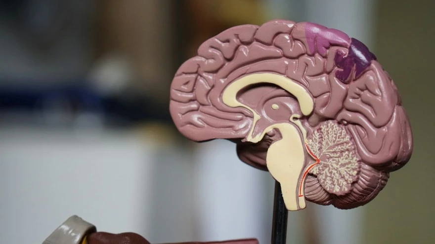
What is a CSF Leak? A cerebrospinal fluid (CSF) leak occurs when the clear, protective fluid surrounding the brain and spinal cord escapes through a tear or hole in the dura mater — the tough outer membrane of the meninges. This clear, colourless fluid acts as a cushion and shock absorber for the central nervous system, delivering essential nutrients and removing waste products. When CSF escapes, it reduces the fluid volume and pressure within the skull and spinal canal, leading to a condition known as spontaneous intracranial hypotension. If left untreated, CSF leaks can cause severe symptoms and potentially life-threatening complications. Types of CSF Leak CSF leaks are classified into two main categories based on their location: Spinal CSF Leak: Occurs along the spinal column due to a tear or hole in the spinal dura mater. Cranial CSF Leak: Develops at the base of the skull, allowing fluid to escape through the nose, ear, or into surrounding tissues. Additionally, CSF leaks can be categorised by their underlying cause: Spontaneous (Primary) CSF Leak: Occurs without an identifiable cause, often associated with structural weaknesses in the dura mater. Nonspontaneous (Secondary) CSF Leak: Results from a known underlying condition, trauma, or medical intervention. CSF Leak Symptoms The symptoms of a CSF leak can vary significantly depending on the location of the leak, making accurate diagnosis challenging. Many patients are initially misdiagnosed with conditions like migraine or fibromyalgia due to overlapping symptoms. The most characteristic symptom of a spinal CSF leak is a severe orthostatic headache that worsens when standing or sitting upright and improves dramatically when lying down. Spinal CSF Leak Symptoms Severe headache that intensifies when upright and improves when lying flat Neck pain or stiffness that may radiate to the shoulders Tinnitus (ringing in the ears) or changes in hearing Dizziness or vertigo, particularly when standing Nausea and vomiting accompanying the headache Visual disturbances, including blurred or double vision Cognitive changes, such as difficulty concentrating or confusion In rare cases, patients may experience a thunderclap headache — a sudden, severe headache that peaks within seconds Pain that worsens with coughing, sneezing, or straining Numbness or tingling sensations in the arms or legs Cranial CSF Leak Symptoms Clear, watery drainage from the nose (rhinorrhea), often from one nostril Clear fluid drainage from the ear (otorrhoea), usually on one side Salty or metallic taste in the mouth due to CSF draining into the throat Increased risk of meningitis due to the opening allowing bacteria to enter the meninges Headache, though typically less severe than in spinal CSF leaks Causes of CSF Leak According to the Spinal CSF Leak Foundation, cerebrospinal fluid (CSF) leaks can occur due to several distinct causes, each affecting the body in different ways and requiring specific diagnostic approaches. Traumatic cause: Head or spinal injuries from accidents, falls, or assaults can cause tears in the dura mater, allowing CSF to leak. Basilar skull fractures are a common traumatic cause of cranial CSF leaks. Medical procedures: Iatrogenic CSF leaks can occur as complications of lumbar punctures, epidural injections, spinal surgeries, or sinus and skull base surgeries. Spontaneous causes: Some CSF leaks develop without apparent trauma or medical intervention. Structural weaknesses in the dura mater, such as meningeal diverticula or abnormal connections between the CSF space and veins (CSF-venous fistulas), can lead to spontaneous leaks. Risk Factors for CSF Leak Connective tissue disorders like Ehlers-Danlos syndrome and Marfan syndrome Previous head, spinal, or sinus surgeries that may have weakened the dura mater Obesity, which may increase intracranial pressure and stress on the dura mater Obstructive sleep apnoea, which can contribute to pressure fluctuations that stress the meninges Intracranial hypertension (increased pressure inside the skull) Irregular skull base anatomy creating structural vulnerabilities Recent spinal or epidural anaesthesia that punctured the dura mater History of head or neck trauma, even if the injury occurred in the past CSF Leak Diagnosis Methods Clinical evaluation and symptom assessment: Doctors carefully take a detailed medical history, focusing on the characteristic orthostatic headache pattern and any recent trauma or procedures. The positional nature of symptoms provides crucial diagnostic clues. Physical examination: Doctors assess for clear fluid drainage from the nose or ears, evaluate neurological function, and may perform tests to check for CSF characteristics in any visible drainage. Pledget study: Small cotton pledgets are placed in the nose to absorb any drainage, which is then tested for beta-2 transferrin or beta-trace protein, markers specific to CSF that confirm the presence of a cranial leak. Imaging studies: Various imaging modalities help locate the site of the leak and assess its severity. This is essential for planning appropriate CSF leak treatment. Intrathecal contrast studies: Contrast material is injected into the spinal canal, and imaging tests are performed to track where the contrast escapes, pinpointing the exact location of spinal leaks. Measurement of opening pressure: During lumbar puncture, the opening pressure of CSF is measured. Low pressure supports the diagnosis of CSF leak causing intracranial hypotension. Imaging & Diagnostic Tests for CSF Leak Imaging tests play a crucial role in diagnosing cerebrospinal fluid (CSF) leaks by helping to localise the site of leakage and assess the extent of the defect. MRI scan of the brain and spine: Reveals signs of intracranial hypotension such as brain sagging, subdural fluid collections, and enhancement of the meninges. MRI myelography can visualise CSF leaks along the spine. CT scan myelography: Computed tomography performed after intrathecal contrast injection provides detailed images showing the precise location of CSF leakage, particularly effective for spinal leaks. Radionuclide cisternography: A nuclear medicine study where a radioactive tracer is injected into the CSF space and tracked over time to identify abnormal CSF flow patterns and leak sites. Here are some of the tests related to CSF leak that you can book at Metropolis Healthcare: Routine Examination (CSF) Beta-trace protein (BTP) test: Detects CSF-specific protein in body fluids to confirm the presence of a CSF leak NMDA receptor antibody test (CSF and serum) CSF Index (CSF and serum) Treatment Options for CSF Leak Conservative measures: For minor leaks or post-lumbar puncture headaches, conservative treatments like bed rest, hydration, and caffeine can help promote healing and alleviate symptoms. Epidural blood patch: A small amount of the patient's own blood is injected into the epidural space, creating a clot that seals the leak. This is often effective for spinal CSF leaks. Epidural patching with fibrin glue: For leaks that don't respond to blood patches, a special glue can be injected to seal the hole or tear in the dura mater. Surgical repair: Persistent or large leaks may require surgical intervention to identify the exact location of the leak and repair the dural defect using sutures, patches, or grafts. Intrathecal saline infusion: In select cases, saline is infused into the spinal canal to temporarily restore CSF pressure and relieve symptoms while natural healing occurs. Conservative Treatment Strict bed rest in a flat position to reduce CSF pressure at the leak site Adequate hydration to promote CSF production and maintain fluid balance Caffeine intake, either orally or intravenously, can constrict cerebral blood vessels and reduce headache severity, aiding symptom relief Stool softeners to prevent straining during bowel movements, which can worsen the leak Analgesic medications to manage headache pain and associated symptoms Close monitoring for signs of improvement or progression Complications of Untreated CSF Leak Chronic debilitating headaches and neurological symptoms that significantly impact quality of life Acquired Chiari malformation, where the brain tissue herniates downward through the foramen magnum due to low CSF pressure Subdural haematoma formation may occur as the brain sags and stretches bridging veins due to low CSF pressure Meningitis or other central nervous system infections due to bacteria entering through the dural defect Cranial nerve palsies, causing symptoms like double vision, facial weakness, or hearing loss Cognitive impairment, memory difficulties, and changes in mental status In severe cases, cerebellar tonsillar descent or herniation can occur, which may be life-threatening Brain haemorrhage may rarely occur from vascular stretching and rupture secondary to brain displacement caused by severe intracranial hypotension Ischaemic stroke or seizures may occur rarely due to altered cerebral perfusion or venous congestion in intracranial hypotension Prevention of CSF Leak While not all CSF leaks are preventable, especially those related to spontaneous causes or predisposing conditions like connective tissue disorders, certain measures can lower the risk of developing a leak. It's essential to wear proper protective equipment and follow safety precautions to reduce the likelihood of head or spinal injuries during high-risk activities. Maintaining a healthy weight can help minimise excessive pressure on the spinal column and meninges. Individuals with obstructive sleep apnoea should seek treatment to manage their condition and avoid sudden pressure changes during sleep. Regular check-ups with a doctor can help identify and address any predisposing factors or early signs of a CSF leak before complications arise. Metropolis Healthcare offers a comprehensive portfolio of over 4,000 tests and profiles, ranging from routine diagnostics to highly specialised tests for cancer, neurological disorders, infectious diseases, and genetic conditions. With a team of qualified phlebotomists who make at-home visits for sample collection and advanced diagnostic labs that process these samples, Metropolis Healthcare ensures reliable results and personalised care. FAQs Can a CSF leak heal on its own? Some CSF leaks, particularly those affecting the spine, may resolve with conservative measures like bed rest. However, many cases require medical intervention or surgical repair. How serious is a CSF leak? CSF leaks can have serious consequences if left untreated, including persistent headaches, infections like meningitis, and potentially permanent neurological damage. What does CSF leak fluid look like? Fluid from a CSF leak is typically clear, watery, and odourless. It may drain from the nose (one nostril) or ear, depending on the leak's location. How long does CSF leak surgery take to heal? Recovery after surgical repair of a CSF leak varies depending on the site and extent of the repair, but most patients notice improvement within weeks, with full recovery typically within one to three months. Full recovery may take one to three months. What is the success rate of CSF leak repair? Surgical treatment of CSF leaks is generally highly successful, with most patients achieving symptom resolution and low rates of recurrence. Can CSF leak cause permanent damage? If a CSF leak is not addressed promptly, it can lead to permanent complications such as hearing loss, neurological deficits, or infection-related damage. References 1. https://my.clevelandclinic.org/health/diseases/16854-cerebrospinal-fluid-csf-leak 2. https://www.mayoclinic.org/diseases-conditions/csf-leak/symptoms-causes/syc-20522246 3. https://spinalcsfleak.org/about-spinal-csf-leaks/causes-of-spinal-csf-leak/ 4. https://www.ncbi.nlm.nih.gov/books/NBK538157/ 5. https://www.hopkinsmedicine.org/health/conditions-and-diseases/cerebrospinal-fluid-csf-leak 6. https://medlineplus.gov/ency/article/001068.htm

Hypoparathyroidism: Symptoms, Diagnosis & Treatment Options
What is Hypoparathyroidism? Hypoparathyroidism is a disorder characterised by deficient secretion or action of parathyroid hormone (PTH), resulting in low calcium (hypocalcaemia) and elevated phosphorus (hyperphosphataemia) levels in the blood. The parathyroid glands — typically four small glands located behind the thyroid — secrete PTH, which helps maintain normal blood calcium levels by stimulating the release of calcium from bones, increasing calcium absorption in the intestines and reducing calcium loss through the kidneys. When the parathyroid glands are damaged, removed, or fail to function properly, PTH levels drop, disrupting the body's ability to regulate calcium balance. This results in hypocalcaemia, which can cause various symptoms related to nerve and muscle function. Understanding Parathyroid Glands and Their Function The parathyroid glands are four small, pea-sized glands located behind the thyroid gland in the neck. They produce and secrete parathyroid hormone (PTH), which plays a vital role in regulating calcium and phosphorus levels in the blood. PTH helps maintain normal blood calcium levels by promoting calcium release from bones, enhancing calcium absorption in the intestines (indirectly through activation of vitamin D), and reducing calcium loss through the kidneys. Proper calcium balance is essential for various bodily functions, including muscle contraction, nerve signalling, and bone health. How Hypoparathyroidism Affects Calcium Balance In hypoparathyroidism, the low PTH levels disrupt the body's ability to maintain normal blood calcium levels, leading to hypocalcaemia. Decreased calcium levels can cause various symptoms, such as tingling sensations, muscle cramps, and spasms. Simultaneously, phosphorus levels rise (hyperphosphataemia) due to reduced excretion by the kidneys, as PTH also regulates phosphorus balance. Prolonged low calcium levels can affect multiple systems, including bones, teeth, hair, skin, and neurological function. Causes of Hypoparathyroidism Surgical removal or damage: The most common cause of hypoparathyroidism is the inadvertent removal or damage of the parathyroid glands during head or neck surgeries, such as thyroidectomy or parathyroidectomy. Autoimmune disorders: In some cases, the body's immune system mistakenly attacks and destroys the parathyroid glands, leading to hypoparathyroidism. This can occur in isolation or as part of a broader autoimmune disease condition, such as autoimmune polyglandular syndrome type 1 (APS-1). Genetic disorders: According to the National Organisation for Rare Disorders, certain rare hereditary conditions, such as DiGeorge syndrome or familial isolated hypoparathyroidism, can cause the parathyroid glands to develop abnormally or function poorly. Radiation therapy: Exposure to radiation in the neck or upper chest area, such as for thyroid or head and neck cancers, can damage the parathyroid glands and cause hypoparathyroidism. Mineral imbalances: Severe magnesium deficiency can impair PTH production and function, resulting in hypoparathyroidism. Idiopathic causes: In some cases, the exact hypoparathyroidism cause remains unknown (idiopathic hypoparathyroidism). Symptoms of Hypoparathyroidism Tingling or numbness in the lips, fingers, and toes (paraesthesia) Muscle cramps and spasms, which may affect the face, hands, feet, or cause difficulty breathing Fatigue and weakness Abdominal pain and nausea Dry, coarse skin and brittle nails Coarse and brittle hair Dental problems, such as delayed tooth development and weak tooth enamel Abnormal heart rhythms (arrhythmias) Emotional changes, including anxiety, depression, and irritability Cataracts (clouding of the eye lens) Painful menstrual periods Seizures (in severe cases) Early Warning Signs Tingling or "pins and needles" sensation in the lips, fingers, or toes Mild muscle cramps or twitching Unusual fatigue or weakness Subtle changes in mood or emotions Diagnosis of Hypoparathyroidism Diagnosing hypoparathyroidism involves a combination of clinical assessment and laboratory tests. Your doctor will consider your medical history, including any recent neck surgeries, family history of endocrine disorders, or autoimmune disease conditions. They will also perform a physical examination to evaluate your hypoparathyroidism symptoms. The diagnosis is confirmed by blood tests showing low calcium levels, high phosphorus levels, and inappropriately low or normal PTH levels. Tests for Hypoparathyroidism Diagnosis Parathyroid Hormone (PTH), Intact assay Parathyroid Panel Test Calcium Profile Vitamin D Plus Profile Calcium Urine 24H Treatment Options for Hypoparathyroidism The primary goal of hypoparathyroidism treatment is to restore and maintain normal blood calcium and phosphorus levels, alleviating symptoms and preventing long-term complications. Treatment options include: Calcium supplements: Oral calcium supplements, such as calcium carbonate or calcium citrate, are prescribed to increase blood calcium levels and manage symptoms. Activated vitamin D supplements: Calcitriol or alfacalcidol, active forms of vitamin D, are used to enhance calcium absorption in the intestines and help maintain normal calcium levels. Thiazide diuretics: In select patients, thiazide diuretics may be used to reduce urinary calcium excretion and help maintain stable calcium levels, particularly in those with hypercalciuria. Magnesium supplementation: If magnesium deficiency is present, magnesium supplements may be necessary to support proper PTH function and calcium regulation. Recombinant human parathyroid hormone (rhPTH 1-84): In patients who do not achieve adequate control with standard therapy, rhPTH 1-84 may be used to replace missing PTH and maintain calcium balance to mimic the action of natural PTH and regulate calcium levels. Dietary adjustments: A diet rich in calcium and magnesium, while limiting phosphorus-rich foods such as dairy, red meat, and processed foods, supports mineral balance. Managing Hypoparathyroidism Long-Term Managing hypoparathyroidism is a lifelong process that involves regular monitoring, medication adjustments, and lifestyle modifications. The goal is to maintain stable blood calcium and phosphorus levels, prevent complications, and ensure optimal quality of life. Patients need to work closely with their healthcare team, including an endocrinologist, to develop an individualised treatment plan. Monitoring and Follow-up Care Periodic blood tests to monitor calcium, phosphorus, and PTH levels Assessment of kidney function through urine calcium and creatinine tests, as well as renal imaging when necessary Monitoring for symptoms of hypocalcaemia or hypercalcaemia, such as neurological and muscular signs Regular eye examinations to check for cataracts Dental check-ups, especially for children, to assess tooth development and enamel health Adjustment of supplement dosages based on laboratory results and clinical response Lifelong follow-up with an endocrinologist to ensure optimal management Complications and Prevention Complications: Kidney stones or nephrocalcinosis (calcium deposits in the kidneys) may occur due to excessive calcium and vitamin D supplementation or prolonged imbalance of calcium and phosphorus levels Seizures and cardiac arrhythmias resulting from severe hypocalcaemia Cataracts, a clouding of the eye lens Dental abnormalities, particularly in children Neuropsychiatric symptoms, such as depression, anxiety, or cognitive impairment Prevention: Careful monitoring of calcium and phosphorus levels, with appropriate adjustments to supplement dosages Early diagnosis and prompt initiation of treatment to prevent long-term complications Patient education on recognizing symptoms of hypocalcaemia or hypercalcaemia and seeking timely medical attention Prognosis and Living with Hypoparathyroidism With appropriate lifelong treatment and regular monitoring, most people with hypoparathyroidism can lead normal, active lives. However, untreated or poorly managed hypoparathyroidism can lead to significant complications and reduced quality of life. Patients need to work closely with their healthcare team, adhere to their treatment plan, and actively participate in their care. Maintaining a balanced diet, staying physically active, and managing stress can also contribute to better overall health and well-being. Metropolis Healthcare, a leading diagnostic laboratory chain with a presence in over 750 towns and cities across India, is committed to providing reliable, patient-centric testing services to support the diagnosis and management of hypoparathyroidism. With a robust network of more than 220 laboratories, 4,600+ service centres, and over 10,000 touchpoints, Metropolis Healthcare offers convenient access to advanced testing. Our commitment to technological innovation, accuracy, and trust has set new benchmarks in the diagnostic industry, enabling healthcare providers to deliver timely, personalised care to patients. FAQs Can hypoparathyroidism be cured? Most cases of hypoparathyroidism cannot be cured, especially when caused by surgical removal or autoimmune destruction of the parathyroid glands. Lifelong treatment is usually necessary, although genetic or transient forms may resolve in rare cases. Is hypoparathyroidism life-threatening? Untreated or poorly managed hypoparathyroidism can be life-threatening due to the risk of severe complications, such as seizures, cardiac arrhythmias, and laryngospasm. However, with proper treatment and monitoring, the risk of life-threatening events is greatly reduced, and patients can lead normal, healthy lives. What is the life expectancy with hypoparathyroidism? With adequate treatment and regular monitoring, the life expectancy for individuals with hypoparathyroidism is generally normal. However, quality of life may be affected by the burden of daily medication, the need for frequent medical check-ups, and the potential for long-term complications if the condition is not well-controlled. Can hypoparathyroidism be reversed? Hypoparathyroidism resulting from reversible causes (such as magnesium deficiency) may improve if the underlying issue is corrected. Permanent cases due to gland removal or genetic causes cannot be reversed. Is hypoparathyroidism hereditary? While most cases of hypoparathyroidism are acquired due to surgery, autoimmune disorders, or other non-genetic causes, some forms of the disorder can be hereditary. Genetic conditions such as familial isolated hypoparathyroidism, DiGeorge syndrome, and autoimmune polyglandular syndrome type 1 (APS-1) can cause hypoparathyroidism. How is hypoparathyroidism different from hyperparathyroidism? While hypoparathyroidism is characterised by low PTH levels, low blood calcium, and high blood phosphorus, hyperparathyroidism presents with high PTH, high calcium, and low phosphorus. Symptoms also differ, with hypoparathyroidism causing muscle cramps, tingling, and seizures, while hyperparathyroidism may lead to bone pain, kidney stones, and abdominal pain. The causes are distinct as well, with hypoparathyroidism often resulting from surgery, autoimmune diseases, or genetic factors, and hyperparathyroidism commonly caused by parathyroid adenomas or hyperplasia. References 1. https://www.mayoclinic.org/diseases-conditions/hypoparathyroidism/symptoms-causes/syc-20355375 2. https://my.clevelandclinic.org/health/diseases/22672-hypoparathyroidism 3. https://rarediseases.org/rare-diseases/hypoparathyroidism/ 4. https://www.ncbi.nlm.nih.gov/books/NBK441899/ 5. https://www.nhs.uk/conditions/hypoparathyroidism/
Urge Incontinence: Bladder Control Tips & Treatment Options
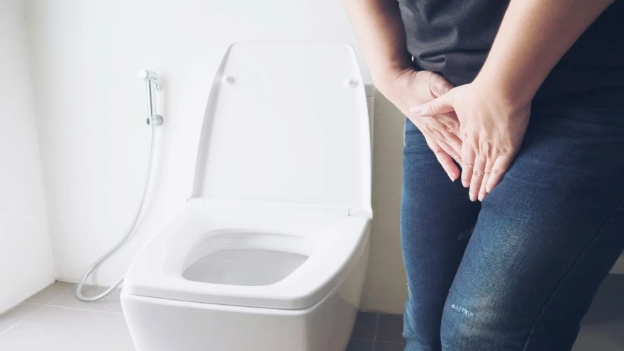
What is Urge Incontinence? Urge incontinence is a form of urinary incontinence characterised by a sudden, compelling urge to urinate that is difficult to defer, often followed by involuntary leakage of urine. When you have urge incontinence, the bladder muscles contract abnormally, sometimes even when the bladder contains only a small amount of urine. This makes it difficult to hold in urine long enough to reach the toilet. Urge incontinence can significantly impact daily activities and quality of life, especially if the episodes are frequent or severe. However, with the right treatment and management strategies, it is possible to regain bladder control and improve your overall well-being. Types of Urinary Incontinence Urge incontinence: Sudden, strong urge to urinate with involuntary leakage Stress incontinence: Leakage that occurs during activities that increase abdominal pressure, such as coughing, sneezing, or lifting Overflow incontinence: Bladder does not empty properly, causing frequent or constant dribbling of urine Mixed incontinence: Combination of symptoms of both urge and stress incontinence Functional incontinence: Inability to get to the toilet in time due to physical or cognitive impairments Urge Incontinence vs Overactive Bladder Urge incontinence and overactive bladder (OAB) are closely related but not identical conditions. Both involve a sudden, compelling need to urinate. However, urge incontinence specifically refers to episodes where this urge leads to involuntary leakage of urine. In contrast, overactive bladder is a broader term describing urinary urgency, often with increased frequency and nocturia (nighttime urination), but it does not always include leakage. It's important to understand the distinction between these two conditions when discussing symptoms with your doctor. Causes of Urge Incontinence Neurological disorders: Conditions such as stroke, multiple sclerosis, Parkinson's disease, and spinal cord injury can disrupt nerve signals between the brain and bladder. Bladder irritation: Infections, stones, inflammation, or tumours in the bladder can cause irritation and lead to urge incontinence. Bladder outlet obstruction: In men, an enlarged prostate (benign prostatic hyperplasia) can cause obstruction and urinary urgency; in women, pelvic organ prolapse may contribute to similar symptoms. Age-related changes: With ageing, the bladder wall may become overactive or less elastic, and pelvic floor muscles may weaken, contributing to urge symptoms. Medications: Certain medications, such as diuretics or sedatives, can increase urinary frequency and urgency. Bladder muscle overactivity: The detrusor muscle in the bladder wall may contract involuntarily, either due to an underlying condition or for unknown reasons. Pelvic surgery or radiation: Previous pelvic surgeries or radiation therapy can affect bladder function. In many cases, no specific cause is found for urge incontinence. However, identifying any underlying factors can help guide treatment decisions. Urge Incontinence Symptoms and Warning Signs Sudden, intense urge to urinate that is difficult to control Involuntary loss of urine immediately following the urge Frequent urination, often more than eight times per day Nocturia, or waking up more than once at night to urinate Leaking urine when hearing or touching running water Large volume of urine lost during leakage episodes Little or no warning before leakage occurs Diagnosis of Urge Incontinence Diagnosing urge incontinence involves a comprehensive evaluation of your medical history, physical examination, and assessment of symptoms. Your doctor will ask about your urinary habits, the onset and severity of symptoms, any associated conditions, medication use, and previous surgeries or neurological issues. During the physical examination, your doctor will evaluate your abdomen, pelvic organs, and nervous system for any abnormalities. They may also ask you to keep a bladder diary. According to the Urology Care Foundation, the bladder diary includes recording your fluid intake, urination times, urine amounts, and any leakage episodes. This information can help identify patterns and triggers for your symptoms. Diagnostic and Imaging Tests for Urge Incontinence To further assess your condition and rule out underlying causes, your doctor may recommend the following tests: Urinalysis and urine culture, such as Urine Routine Test (Urine R/M Test), to check for urinary tract infection or blood in the urine Urine Culture and Sensitivity Test to identify urinary tract infections (UTIs) and the specific bacteria causing infection Post-void residual measurement to assess how well your bladder empties, often using ultrasound Urodynamic studies to measure bladder pressure and function during filling and emptying Cystoscopy, which involves inserting a thin, lighted tube into the urethra to directly visualise the bladder for any structural abnormalities Imaging tests such as renal and bladder ultrasound, and in select cases MRI, may be used to evaluate structural or neurological causes Treatment Options for Urge Incontinence Behavioural therapies and bladder training: This involves scheduled voiding patterns to gradually increase the time between bathroom visits, helping retrain the bladder to hold urine longer. Bladder training teaches you to delay urination when you feel the urge, starting with small delays and progressively extending them. Pelvic floor muscle exercises (Kegel exercises): A decline in oestrogen levels after menopause can reduce pelvic floor muscle tone and urogenital tissue support, contributing to urinary symptoms such as urgency and leakage. Performing targeted exercises to strengthen these muscles can help enhance bladder control. Biofeedback and pelvic floor physical therapy: Working with specialised physical therapists who use biofeedback devices helps you identify and properly engage your pelvic floor muscles. This approach provides real-time feedback to ensure exercises are performed correctly. Pharmacological treatment: Antimuscarinic medications (also called anticholinergics) reduce involuntary bladder contractions by blocking acetylcholine at muscarinic receptors, helping the bladder hold more urine. Beta-3 agonists represent another medication class that helps the bladder hold more urine. Neuromodulation therapies: These include sacral nerve stimulation and percutaneous tibial nerve stimulation, which use electrical impulses to modify nerve signals between the bladder and brain, reducing urgency and frequency. Botulinum toxin (onabotulinumtoxinA) injections: Administered directly into the detrusor muscle, Botox reduces involuntary contractions and increases bladder capacity for several months. Surgical interventions: For severe cases unresponsive to other treatments, surgical options like bladder augmentation (enlarging the bladder) or urinary diversion procedures may be considered. Lifestyle Changes for Better Bladder Control Maintain a healthy weight to reduce pressure on the bladder and pelvic floor. Implement timed voiding schedules, urinating every 2-4 hours rather than waiting for urgency. Practise double voiding, waiting a few moments after urinating to attempt emptying the bladder again. Manage fluid intake, avoiding large amounts at once and limiting fluids before bedtime. Avoid known bladder irritants like caffeine, alcohol, carbonated drinks, artificial sweeteners, and spicy or acidic foods. Quit smoking to reduce bladder irritation and chronic cough. Treat constipation promptly to avoid pressure on the bladder. Practise stress management techniques like deep breathing, meditation, or yoga. Foods and Drinks That Trigger Urge Incontinence What you eat and drink can directly impact bladder function. Certain foods and beverages are known to irritate the bladder lining, increasing urinary urgency and frequency. Try eliminating or cutting back on these common culprits: Caffeinated drinks like coffee, tea, sodas, and energy drinks Alcoholic beverages, including beer, wine, and spirits Carbonated drinks, even if caffeine-free Citrus fruits and juices like oranges, grapefruits, lemons, and limes Tomato-based products, including sauces, soups, and ketchup Spicy foods containing hot peppers or curry Artificial sweeteners such as aspartame and saccharin Chocolate, which contains caffeine and other stimulants Acidic foods like vinegar, pickles, and citrus fruits Instead, opt for bladder-friendly alternatives like water, herbal teas, milk, non-citrus fruits, whole grains, and lean proteins. Keeping a food diary can help you identify potential triggers to avoid. Medications for Urge Incontinence Anticholinergic medicines are the mainstay of pharmacological treatment for overactive bladder. They work by blocking acetylcholine, a neurotransmitter that causes the detrusor muscle to contract. This allows the bladder to relax and hold more urine, reducing episodes of urgency and frequency. Commonly prescribed anticholinergics include oxybutynin, tolterodine, darifenacin, and solifenacin. While generally well-tolerated, anticholinergic side effects may include dry mouth, constipation, blurred vision, and drowsiness, especially in older adults. Extended-release formulations and transdermal patches often have fewer side effects than immediate-release pills. Mirabegron, a beta-3 adrenergic agonist, relaxes the detrusor muscle via adrenergic stimulation, improving bladder storage with fewer cognitive or dry mouth side effects compared to antimuscarinics. It may be used alone or in combination with an anticholinergic. Advanced Treatment Options For individuals who don't respond to conservative measures or can't tolerate medications, several advanced therapies are available: Sacral neuromodulation (InterStim): A surgically implanted device sends electrical pulses to the sacral nerves, modulating signals between the bladder and brain. Percutaneous tibial nerve stimulation (PTNS): A minimally invasive procedure that stimulates the tibial nerve near the ankle to regulate bladder activity. Botox injections: Injecting botulinum toxin into the bladder muscle temporarily paralyses it, reducing contractions and increasing capacity for 6-12 months. Bladder augmentation surgery: Enlarging the bladder with a segment of intestine can increase capacity for those with severe symptoms. Urinary diversion: Creating an alternate pathway for urine storage and elimination may be a last resort for intractable cases. Prevention of Urge Incontinence While some urge incontinence risk factors like age and genetics are beyond your control, you can take proactive steps to prevent bladder control problems: Maintain a healthy body mass index (BMI) to minimise pressure on the pelvic floor. Perform pelvic floor exercises regularly to optimise muscle strength before issues arise. Avoid chronic constipation by consuming adequate fibre and water. Limit irritating foods and drinks to decrease bladder sensitivity over time. Properly manage chronic conditions like diabetes and neurological disorders. Don't smoke, as it contributes to urge incontinence in several ways. Avoid frequent ‘just-in-case’ urination, as it can train the bladder to signal urgency at lower volumes. Stay hydrated with water to avoid concentrated urine that irritates the bladder. Promptly treat UTIs to prevent bladder irritation and damage. Living with Urge Incontinence Living with urge incontinence requires adaptation and comprehensive management strategies, but many individuals successfully maintain active, fulfilling lives with appropriate urge incontinence treatment and support. The condition can significantly impact quality of life, leading to social isolation, anxiety about public outings, sleep disruption from nocturia, and intimate relationship challenges. However, by working closely with doctors, implementing lifestyle modifications, and adhering to individualised treatment plans, most people can effectively control their urge incontinence symptoms and minimise the impact on daily activities. Seeking support from loved ones, counsellors, or support groups can provide valuable emotional resources and practical tips for navigating the challenges of living with urge incontinence. Remember, you are not alone in this journey, and with the right tools and mindset, it is possible to regain control and confidence. Metropolis Healthcare offers a comprehensive portfolio of more than 4,000 tests and profiles, ranging from routine diagnostics to highly specialised tests for cancer, neurological disorders, infectious diseases, and genetic conditions. Our experienced phlebotomists offer convenient at-home sample collection, ensuring your comfort and privacy. Test reports are promptly delivered via email and the user-friendly Metropolis Healthcare App, empowering you to make informed health choices. FAQs Can urge incontinence be cured? While there is no definitive cure for urge incontinence, many individuals experience significant improvement or complete symptom resolution with a combination of lifestyle modifications, behavioural therapies, medications, and advanced treatment options. What is the success rate of urge incontinence treatment? The success rate of urge incontinence treatment varies depending on the individual and the specific therapies employed. However, studies have shown that behavioural therapies and pelvic floor exercises can yield up to 80% improvement in symptoms, while medications can reduce urgency episodes by 50-70%. How long does it take for urge incontinence treatment to work? The timeline for seeing results from urge incontinence treatment depends on the specific therapies used. Behavioural modifications and pelvic floor exercises may show improvement within a few weeks to months, while medications typically require 4-12 weeks to reach full effectiveness. Advanced therapies such as neuromodulation or Botox injections may begin showing symptom relief within days to weeks, depending on individual response. What foods should I avoid with urge incontinence? To minimise bladder irritation and urgency, it's best to avoid or limit caffeine, alcohol, carbonated beverages, citrus fruits and juices, tomato-based products, spicy foods, artificial sweeteners, chocolate, and acidic foods. Is urge incontinence worse at night? Urge incontinence can be particularly bothersome at night, as the sudden need to urinate can disrupt sleep and lead to bedwetting. Nocturia, or frequent nighttime urination, is a common symptom of overactive bladder. Limiting fluid intake 2-3 hours before bedtime and using absorbent products can help manage nighttime symptoms. Can stress cause urge incontinence to worsen? Yes, stress and anxiety can exacerbate urge incontinence symptoms by increasing muscle tension and stimulating the bladder. Practising stress management techniques like deep breathing, meditation, or yoga can help reduce the impact of stress on bladder control. References 1. https://my.clevelandclinic.org/health/diseases/22161-urge-incontinence 2. https://medlineplus.gov/ency/article/001270.htm 3. https://www.urologyhealth.org/urology-a-z/u/urinary-incontinence 4. https://www.ncbi.nlm.nih.gov/books/NBK563172/ 5. https://www.nhs.uk/conditions/urinary-incontinence/surgery/

Brown Bread vs White Bread: Health Differences You Should Know
What is Brown Bread? Brown bread is commonly made from whole wheat flour, which includes all parts of the wheat grain — the bran, germ, and endosperm — unless it has been refined or coloured. This preserves the natural fibre, vitamins, and minerals found in wheat. However, not all brown bread on the market is 100% whole wheat. Some commercial varieties are made from refined flour and coloured with additives like caramel or molasses to appear healthier. What is White Bread? White bread, on the other hand, is made from refined wheat flour, where the bran and germ are removed during processing, resulting in a softer texture and lighter colour. According to the Whole Grains Council, refining whole wheat flour to make white flour removes the bran and germ, stripping away most of the fibre, vitamins (including E and B6), minerals (such as potassium and magnesium), and phytonutrients. Brown Bread vs White Bread: Key Differences Feature Brown Bread White Bread Main Ingredient Whole wheat flour (contains bran & germ) Refined wheat flour (endosperm only) Fibre Content High Low Vitamin & Mineral Content Higher in naturally occurring B vitamins, magnesium, zinc, and folate (B9) Lower (some nutrients may be artificially added) Glycaemic Index Lower (slower blood sugar impact) Higher (faster blood sugar spike) Calories per Slice ~75 - 80 ~77- 80 Texture Denser, chewier Softer, lighter Common Additives Sometimes coloured with caramel/molasses May be bleached, fortified Nutritional Value of Brown Bread High in dietary fibre, which aids digestion and promotes satiety Contains B vitamins (B6, folic acid), vitamin E, and minerals like magnesium, zinc, copper, and manganese Rich in antioxidants due to intact wheat germ and bran Has a lower glycaemic index than white bread, leading to steadier post-meal blood sugar levels Slightly fewer calories than white bread Nutritional Value of White Bread Low in fibre due to removal of bran and germ during processing May be fortified with select vitamins and minerals but often lacks natural nutrient density Higher glycaemic index, causing quicker spikes in blood sugar Typically higher in calories per slice than brown bread, though the difference is minimal Contains more refined starch and less protein than whole-grain breads Health Benefits of Brown Bread Supports healthy digestion due to high fibre content Helps regulate blood sugar levels, beneficial for diabetes management Whole-grain brown bread can support weight management by promoting fullness (satiety) and potentially reducing overall calorie intake when consumed as part of a balanced diet Reduces risk of heart disease through improved cholesterol regulation Provides a broader range of essential vitamins and minerals Health Benefits of White Bread Easily digestible for individuals with certain digestive issues Provides a quick energy source due to high refined carbohydrate content Sometimes fortified with iron and B vitamins to address common nutrient deficiencies, though naturally occurring nutrients are lower Soft texture may be preferred by children and older adults Health Risks of Brown Bread Many commercial ‘brown breads’ are coloured with caramel or molasses and may contain little or no whole grain, offering few added health benefits Those with gluten intolerance or wheat allergy should avoid both brown and white wheat-based breads May be denser and less palatable for some, potentially limiting intake of fibre if not consumed regularly Health Risks of White Bread Low fibre can lead to poor digestive health and increased risk of constipation High glycaemic index may contribute to spikes in blood sugar, increasing risk of type 2 diabetes and heart disease for frequent consumers Associated with weight gain when consumed in excess due to low satiety and high refined carbohydrate content Fewer natural vitamins and minerals compared to whole grain options Brown Bread vs White Bread for Weight Loss When it comes to brown bread vs white bread calories, the difference is minimal. However, brown bread is generally preferable for weight loss because it is higher in fibre, which helps you feel full longer and may reduce overall calorie intake. Brown bread also leads to slower rises in blood sugar compared to white bread, supporting better appetite control and metabolic health. However, simply switching to brown bread is not sufficient for weight loss; total diet quality, portion size, and physical activity all play critical roles. Consider these points when choosing bread for weight management: Brown bread increases satiety due to fibre A lower glycaemic index helps manage hunger and energy White bread can cause rapid increases and decreases in blood sugar (glycaemic variability), especially when eaten without protein or fibre Portion control and physical activity remain essential Which Bread is Better for Diabetes? For individuals with diabetes, brown bread, specifically made from whole wheat or whole grains, is a better choice due to its lower glycaemic index and higher fibre content. These properties help stabilise blood sugar levels, reducing spikes after meals. In contrast, white bread, made from refined flour, is digested quickly and can cause rapid increases in blood sugar, which is not ideal for diabetes management. Brown Bread vs White Bread: Taste & Texture In terms of taste and texture, brown bread tends to be denser and chewier, with a more pronounced, slightly nutty flavour due to the presence of bran and germ. White bread is softer, lighter, and milder in taste, making it more appealing to some people, especially children, who prefer a less coarse texture. Ultimately, preferences for taste and texture are subjective and can influence bread choice regardless of nutritional differences. Hidden Truth: Is All Brown Bread Really Healthy? It's important to note that not all brown bread is truly healthy. Many commercial varieties are made with refined flour and coloured with caramel or molasses to mimic whole grain bread, offering little more nutrition than white bread. To ensure you're getting the health benefits of brown bread, check the ingredients list for "100% whole wheat" or "whole grain" as the primary ingredient and avoid breads labelled as "enriched" or with added colours. Brown Bread vs White Bread: Expert Dietitian Advice Expert dietitians recommend choosing breads labelled as "100% whole wheat" or "whole grain" for maximum nutritional benefit, emphasising the importance of fibre, vitamins, and minerals found naturally in whole grains. They advise being cautious of misleading labels and suggest reading ingredient lists carefully to avoid breads that use mostly refined flour with added colouring or sugar. Moderation in overall bread consumption is also key, even with whole grain varieties. How to Choose the Right Bread at the Store Check the ingredient list thoroughly and look for "100% whole wheat" or "whole grain" as the first ingredient Avoid breads with caramel colour or excessive additives Compare fibre content; higher fibre means more whole grain Look for minimal added sugars and sodium Ignore colour, as brown does not always mean whole grain Check for fortification if you need specific nutrients (like iron or B vitamins) Consider your dietary restrictions (e.g., gluten-free, low-carb) Choose products with fewer ingredients for less processing Brown Bread vs White Bread: Final Verdict When comparing brown bread vs white bread, brown bread (especially 100% whole wheat or whole grain) is generally healthier due to its higher fibre, vitamin, and mineral content, and lower glycaemic index. However, not all brown breads are created equal, so careful label reading is essential. White bread can be included in a balanced diet in moderation but should not be the primary grain source for most adults. Metropolis Healthcare has an extensive network spanning over 220 laboratories and more than 4600 service centres across 750+ towns in India, with over 10,000 consumer touchpoints. With a team of skilled blood collection technicians who make convenient at-home visits, Metropolis ensures convenience and comfort for patients. Our state-of-the-art diagnostic labs process samples efficiently, and test reports are securely shared online via email and the user-friendly Metropolis Healthcare App. FAQs Is brown bread really healthier than white bread? Yes, brown bread made from whole wheat is higher in fibre and nutrients, making it healthier than white bread made from refined flour. Can white bread cause weight gain? Eating too much white bread, which is low in fibre and high in refined carbs, can contribute to weight gain if it's not balanced with an overall healthy diet and physical activity. Which bread is better for diabetics – brown or white? Whole grain brown bread is a better option for people with diabetes because it has a lower glycaemic index and is digested more slowly, helping to control blood sugar levels. Does brown bread have less calories than white bread? The brown bread vs white bread calories difference is minimal, with brown bread having slightly fewer calories per slice on average. However, brown bread is more filling due to its higher fibre content. What is the healthiest bread option apart from brown bread? Other healthy bread options include 100% whole grain breads made from oats, rye, barley, quinoa, or sprouted grains, and sourdough made with whole grain flours, as well as sourdough bread, which is more easily digestible. Can children eat brown bread daily? Yes, children can eat whole grain brown bread daily as part of a balanced diet. It provides them with essential nutrients and fibre for healthy growth and development. Is all brown bread whole wheat? No, not all brown bread is whole wheat. Some commercial brown breads are made from refined flour with added colouring to appear healthier. Always check labels for "100% whole wheat" as the first ingredient. How much bread should you eat in a day? The amount of bread you should eat each day depends on factors such as your age, sex, and activity level. In general, adults are advised to consume about 6–10 servings (ounce-equivalents) of grains per day, with at least half of these coming from whole-grain sources. One regular slice of bread typically counts as one serving. References 1. https://nutritionsource.hsph.harvard.edu/what-should-you-eat/whole-grains/ 2. https://www.mayoclinic.org/healthy-lifestyle/nutrition-and-healthy-eating/in-depth/whole-grains/art-20047826 3. https://wholegrainscouncil.org/blog/2018/01/myths-busted-why-white-bread-not-just-healthy-whole-grain-bread 4. https://pmc.ncbi.nlm.nih.gov/articles/PMC8181512/ 5. https://health.clevelandclinic.org/bread-best-whole-grain-multigrain-whole-wheat 6. https://myplate4chatbot.stg.platform.usda.gov/eat-healthy/grains

Pelvic Organ Prolapse: Symptoms, Grading & Way Forward
What is Pelvic Organ Prolapse? Pelvic organ prolapse (POP) occurs when one or more of the pelvic organs (such as the bladder, uterus, rectum, or small intestine) descend from their normal position and bulge into or out of the vagina. This happens due to weakened muscles or ligaments that support the pelvic floor. Factors like childbirth, ageing, menopause, chronic pressure (from obesity or constipation), or pelvic surgery can contribute to this weakening. According to the National Health Service (NHS), pelvic organ prolapse is common in women over 50. POP can affect anyone with a vagina, and severity ranges from mild cases that often cause no symptoms to more advanced prolapse that leads to significant discomfort and functional problems. Types of Pelvic Organ Prolapse There are several types of pelvic organ prolapse, depending on which organ is affected: Cystocele (bladder prolapse): The bladder bulges into the front wall of the vagina. This can cause discomfort, urinary incontinence, or a feeling of pressure in the pelvic area. Women may notice difficulty emptying their bladder completely or frequent urinary tract infections. Uterine prolapse: The uterus descends into the vaginal canal. This may cause a sensation of heaviness or pulling in the pelvis and sometimes visible tissue protruding from the vaginal opening. Severe cases can interfere with sexual activity and cause urinary or bowel problems. Rectocele: The rectum bulges into the posterior vaginal wall, leading to difficulty with bowel movements such as constipation or the need for vaginal pressure support (splinting). Enterocoele (small bowel prolapse): A herniation of the small intestine into the upper posterior vaginal wall, often through a weakened vaginal apex. It often causes pelvic pressure or a pulling sensation, especially during activities like lifting or straining. Enteroceles can sometimes contribute to vaginal fullness or discomfort deep inside the pelvis. Vaginal vault prolapse: Descent of the upper portion of the vaginal canal, typically following hysterectomy, due to loss of apical support. This occurs because the support structures for the vaginal apex weaken or fail. It can cause a sensation of vaginal bulging, discomfort, and sometimes difficulty with intercourse. Symptoms of Pelvic Organ Prolapse Feeling or seeing a bulge in or out of the vagina Sensation of heaviness, pressure, or fullness in the pelvis or vagina Low backache or pelvic pain Discomfort or pain during intercourse Urinary problems, such as leakage, slow stream, incomplete emptying, frequency, or urinary tract infections Bowel symptoms, like constipation, incomplete evacuation, or accidental leakage Difficulty inserting a tampon Spotting or vaginal bleeding Worsening of symptoms when standing, during physical activity, or at the end of the day It's important to note that symptom severity doesn't always correlate with the degree of prolapse. Even mild cases can cause significant discomfort, while advanced prolapse may present with subtle symptoms. Vaginal Bulge and Pressure One of the most noticeable symptoms of pelvic organ prolapse is a sensation of pressure, fullness, or heaviness in the pelvis or vagina. Many women describe this feeling as a dragging or pulling sensation, as if something is "falling out" of their vagina. This sensation may be accompanied by a feeling of fullness, heaviness, or pressure in the pelvic region, which can worsen after standing for long periods, during physical activity, or as the day progresses. In more advanced cases, the bulging tissue may be visible at or beyond the vaginal opening. Some women notice or feel the bulge during bathing or with a mirror. This symptom can be distressing and impact daily activities, self-image, and sexual function. Urinary and Bowel Changes Pelvic organ prolapse can lead to changes in urinary and bowel functions, which can be frustrating and embarrassing. Common urinary symptoms include: Leakage of urine (stress incontinence), especially when coughing, laughing, or exercising Difficulty starting urination or a slow urine stream Feeling of incomplete bladder emptying or needing to urinate more frequently Urgency: a sudden, strong need to urinate Increased risk of urinary tract infections Prolapse can also affect bowel movements, causing: Difficulty with bowel movements, including constipation or needing to strain Sensation of incomplete evacuation Accidental bowel leakage or incontinence These symptoms occur because the prolapsed organs can press against the bladder or rectum, disrupting their normal functions. In some cases, women may need to manually support the prolapsed tissue to initiate or complete urination or bowel movements. Untreated severe POP can occasionally lead to complications such as recurrent urinary tract infections, pelvic discomfort, or hydronephrosis due to urinary obstruction. Grading & Stages of Pelvic Organ Prolapse (Stage I–IV Classification) The severity of pelvic organ prolapse is assessed using a grading system called the Pelvic Organ Prolapse Quantification (POP-Q) system. This system assigns a stage from 0 to IV based on precise measurements taken during a pelvic exam. The grading helps doctors objectively describe the extent of the prolapse and guide treatment decisions. The stages of pelvic organ prolapse are classified as follows: Stage 0: No prolapse; organs are in their normal position. Stage I: The most descended portion of the prolapse is more than 1 cm above the hymen (the vaginal opening). Stage II: The most descended portion is within 1 cm above or below the hymen. Stage III: The most descended portion is more than 1 cm below the hymen but not completely outside the vagina. Stage IV: Complete eversion; the vaginal walls are fully everted and the uterus (if present) may protrude outside the vaginal opening (procidentia). It's important to note that: Mild prolapse (Stages I–II) is often asymptomatic or associated with mild symptoms, as the organs are still within the vagina. Moderate prolapse (Stage III) involves organs descending beyond the hymen but not completely outside the vagina; symptoms are usually more pronounced at this stage. Severe prolapse (Stage IV) is rare but can cause significant discomfort and functional impairment, as the vagina is completely turned inside out. If you suspect you have pelvic organ prolapse, it's essential to consult a doctor for a proper diagnosis and treatment plan. Your doctor may recommend conservative measures such as pelvic floor exercises, pessaries, or lifestyle changes for mild to moderate cases. More severe cases may require surgical intervention to restore the organs to their normal position and improve symptoms. Causes of Pelvic Organ Prolapse Pelvic organ prolapse results from a combination of factors that weaken or damage the pelvic floor support structures to compromise the structural integrity of the pelvic floor. Vaginal childbirth is the most significant risk factor, particularly when deliveries involve prolonged labour, multiple births, forceps or vacuum assistance, or large birth weight infants. As women age, their pelvic tissues naturally weaken, and oestrogen deficiency after menopause further contributes to tissue atrophy and loss of collagen strength. Chronic conditions that increase intra-abdominal pressure, such as chronic cough from pulmonary disease, chronic constipation requiring repetitive straining, and obesity, place sustained stress on pelvic support structures. Previous pelvic surgeries, particularly hysterectomy, can disrupt the normal support mechanisms of the vaginal apex. Genetic factors influencing connective tissue composition, like collagen disorders such as Ehlers-Danlos syndrome and Marfan syndrome, also predispose some women to developing prolapse. Heavy lifting, either occupational or recreational, and conditions causing chronic ascites further contribute to the progressive weakening of pelvic support over time. Dietary & Lifestyle Modifications Making specific dietary and lifestyle changes can significantly reduce symptoms and prevent the progression of pelvic organ prolapse. Maintaining a healthy body weight through balanced nutrition alleviates excessive pressure on the pelvic floor, as obesity substantially increases the risk of prolapse development and worsening. Consuming a high-fibre diet prevents constipation and reduces the need for straining during bowel movements, which repeatedly stresses pelvic support structures. Adequate hydration, typically eight to ten glasses of water daily, supports regular bowel function and helps maintain tissue health. Women should avoid heavy lifting whenever possible or learn proper lifting techniques that engage core muscles rather than bearing down with the pelvic floor. Treating chronic cough through smoking cessation and managing respiratory conditions prevents repetitive increases in intra-abdominal pressure. For postmenopausal women, topical vaginal oestrogen therapy may help maintain local tissue elasticity and reduce vaginal atrophy. Regular pelvic floor exercises, commonly known as Kegel exercises, strengthen the muscles supporting the pelvic organs and can prevent mild prolapse from progressing. These lifestyle modifications work synergistically to reduce mechanical stress on already compromised pelvic support structures. Diagnosis and Assessment Diagnosing pelvic organ prolapse involves a thorough evaluation of your medical history, focusing on symptoms like a feeling of vaginal fullness, urinary problems, and bowel changes. Your doctor will perform a pelvic exam, assessing the degree of prolapse and identifying the affected organs. Standardised grading systems, such as the Pelvic Organ Prolapse Quantification (POP-Q) system or the Baden-Walker halfway system, are used to objectively stage the prolapse. These systems help your doctor determine the severity and guide treatment decisions. Examinations and Tests to Diagnose Pelvic Organ Prolapse Pelvic examination using a speculum to visualise all vaginal compartments and identify prolapsing organs POP-Q staging assessment for objective, reproducible staging of prolapse severity Pelvic floor muscle strength assessment to guide individualised management planning Urinalysis, such as Urine Routine Test (Urine R/M Test), to check for infection, particularly when bladder symptoms are present Bladder scan and urodynamic studies to assess bladder function and capacity when urinary symptoms are reported Examination in multiple positions, such as lying on the side or standing upright, to fully appreciate prolapse extent Digital vaginal examination to palpate for lumps and rule out differential diagnoses Treatment Options & Way Forward Pelvic organ prolapse treatment must be individualised based on prolapse severity, symptom burden, patient age, general health status, sexual activity, and family planning desires. Pelvic organ prolapse should only be considered requiring intervention if it causes bothersome pressure symptoms, affects sexual function, or disrupts normal lower urinary tract or bowel function. For women with Stage 1 prolapse who typically have mild or no symptoms, a "wait and see" approach with observation is appropriate, as prolapse is not life-threatening. Conservative management includes pelvic floor muscle training (Kegel exercises) performed regularly to strengthen supportive muscles, which can be enhanced through pelvic floor physical therapy with biofeedback techniques. Vaginal pessaries—removable silicone or plastic devices inserted into the vagina—provide mechanical support for prolapsed organs and represent an excellent non-surgical option for women who prefer to avoid surgery, have medical contraindications to surgery, or wish to delay surgical intervention. For postmenopausal women, vaginal oestrogen therapy may improve tissue quality and reduce prolapse-related symptoms. Surgical intervention becomes appropriate when conservative measures fail to adequately control symptoms or when prolapse significantly impairs quality of life. Surgical options include reconstructive procedures that repair and restore normal anatomy using the patient's own tissues or, in select cases, synthetic mesh materials, though mesh use has become more restricted due to potential complications. Obliterative procedures, which close part or all of the vaginal canal, provide a definitive treatment option for women who are not sexually active and prefer a lower-risk surgical approach. Preventive Strategies Perform regular pelvic floor exercises (Kegel exercises) to strengthen the muscles supporting pelvic organs and prevent mild prolapse from developing or progressing Maintain a healthy body weight to reduce chronic pressure on the pelvic floor and decrease prolapse risk Prevent and treat constipation by consuming a high-fiber diet with adequate hydration to avoid straining during bowel movements Avoid heavy lifting or use proper body mechanics, engaging core muscles and avoiding bearing down with the pelvic floor Treat chronic cough promptly to prevent repetitive increases in intra-abdominal pressure that weaken pelvic support structures Consider vaginal oestrogen therapy for postmenopausal women to maintain vaginal and pelvic tissue integrity Schedule regular gynaecological check-ups to monitor pelvic health and address any concerns promptly At Metropolis Healthcare, we understand the importance of early detection and personalised care in managing pelvic floor disorders. Our team of skilled phlebotomists offers convenient at-home sample collection services, ensuring your comfort and privacy. With a comprehensive portfolio of over 4,000 tests and profiles, ranging from routine diagnostics to highly specialised tests for cancer, neurological disorders, infectious diseases, and genetic conditions, Metropolis is committed to providing accurate, reliable results to guide your healthcare journey. FAQs What are the first signs of pelvic organ prolapse? Early symptoms of pelvic organ prolapse may include a feeling of heaviness or pressure in the vagina, a visible or palpable bulge, urinary leakage or difficulty emptying the bladder, constipation or incomplete bowel emptying, and discomfort during intercourse. Can pelvic organ prolapse fix itself? While mild cases of pelvic organ prolapse may improve with conservative measures like pelvic floor exercises and lifestyle changes, the condition usually does not resolve completely on its own. Seeking medical advice is important for proper diagnosis and treatment. What is the best treatment for pelvic organ prolapse? The best pelvic organ prolapse treatment varies depending on the severity of the prolapse and the individual's symptoms and preferences. Options range from lifestyle modifications and pessary devices to surgical repair. Personalised care is key for optimal results. Is pelvic organ prolapse painful? Some women with pelvic organ prolapse may experience discomfort, pelvic pressure, lower back pain, or pain during intercourse. However, others may have no pain and only notice a bulge or a feeling of fullness in the vagina. Can pelvic organ prolapse come back after surgery? While surgical repair can effectively treat pelvic organ prolapse, there is a risk of recurrence, especially if contributing factors like obesity or chronic straining persist. Regular follow-up care and preventive strategies can help reduce the likelihood of prolapse returning. How can I prevent pelvic organ prolapse? To lower your risk of developing pelvic organ prolapse, maintain a healthy weight, perform regular pelvic floor exercises, eat a fibre-rich diet to prevent constipation, avoid heavy lifting and high-impact activities, treat chronic cough promptly, and seek early medical care for urinary or bowel symptoms. References 1. https://my.clevelandclinic.org/health/diseases/24046-pelvic-organ-prolapse 2. https://www.nhs.uk/conditions/pelvic-organ-prolapse/ 3. https://www.mayoclinic.org/diseases-conditions/pelvic-organ-prolapse/symptoms-causes/syc-20360557 4. https://www.ncbi.nlm.nih.gov/books/NBK563229/ 5. https://www.betterhealth.vic.gov.au/health/conditionsandtreatments/bladder-prolapse

Hydrogen Breath Test: What It Diagnoses & How to Prepare
What Is the Hydrogen Breath Test? The hydrogen–methane breath test is a simple, non-invasive diagnostic tool that measures the concentration of hydrogen and methane gases in your exhaled breath to detect various gastrointestinal disorders. The test is based on the principle that when certain sugars or carbohydrates are not properly digested and absorbed in the small intestine, they reach the colon, where bacteria ferment them, producing hydrogen gas as a byproduct. This hydrogen is then absorbed into the bloodstream, transported to the lungs, and exhaled in your breath, where it can be measured using specialised equipment. By analysing the pattern and levels of hydrogen and methane gases in your breath samples over a period of time, doctors can identify specific digestive issues and guide treatment decisions. Conditions Diagnosed by the Hydrogen Breath Test Lactose Intolerance: This occurs when the body lacks sufficient lactase, the enzyme needed to digest lactose (milk sugar). Undigested lactose ferments in the colon, causing symptoms like bloating, diarrhoea, and abdominal pain. Fructose Malabsorption: Similar to lactose intolerance, fructose malabsorption involves the inability to properly absorb fructose (fruit sugar), leading to fermentation by gut bacteria and digestive symptoms. Small Intestinal Bacterial Overgrowth (SIBO): SIBO is characterised by an abnormal increase in the number of bacteria in the small intestine, which can interfere with normal digestion and cause bloating, diarrhoea, and malnutrition. Irritable Bowel Syndrome (IBS): The hydrogen–methane breath test may help identify overlapping conditions such as SIBO or carbohydrate malabsorption that can mimic or contribute to IBS-like symptoms. Additionally, the test can be used to evaluate less common carbohydrate intolerances, such as sucrose or sorbitol malabsorption, and to assess orocecal transit time. How the Hydrogen Breath Test Works Baseline Measurement: You'll provide an initial breath sample by exhaling into a collection bag to establish your baseline hydrogen levels before consuming the test sugar solution. Sugar Solution Consumption: You’ll drink a solution containing a specific type of carbohydrate substrate (such as lactose, lactulose, fructose, or glucose) that will challenge your digestive system. Bacterial Fermentation: If the sugar is not properly digested in the small intestine, it travels to the colon (or encounters bacteria in the small intestine in cases of SIBO), where anaerobic bacteria ferment the unabsorbed carbohydrates. Hydrogen Production and Absorption: The bacterial fermentation process produces hydrogen gas, which is absorbed through the intestinal walls into the bloodstream. Transport to Lungs: The absorbed hydrogen travels through the bloodstream to the lungs, where it diffuses into the air spaces. Breath Collection at Intervals: You'll provide Breath samples every 15 to 20 minutes over a period of two to three hours, depending on the substrate used as your body digests the sugar solution. Measurement and Analysis: Each breath sample is analysed to measure the hydrogen concentration in parts per million (ppm). Types of Substrates Used in Testing Lactose: Used to diagnose lactose intolerance Fructose: Used to diagnose fructose malabsorption Lactulose: Used to assess small intestinal transit time and, indirectly, to screen for SIBO Glucose: Also used to diagnose SIBO Sucrose: Used to detect sucrose intolerance Sorbitol: Used to identify sorbitol intolerance Preparing for Your Hydrogen Breath Test Fast for 12 hours before the test, avoiding all food and beverages except water. Avoid high-fibre foods, dairy products, and fermentable carbohydrates (such as beans, onions, and whole grains) the day before your test, as these can affect bacterial fermentation patterns. Inform your doctor about any medications or supplements you're taking, as some may need to be temporarily discontinued before the test (such as antibiotics, probiotics, or laxatives), according to the NHS Trust. Refrain from smoking or engaging in strenuous physical activity for at least 2 hours before and during the test, as these can influence hydrogen production and breath measurements. On the morning of the test, brush your teeth without using toothpaste or mouthwash, as these products may contain sugars that could interfere with results. Which Foods and Medications to Avoid Before the Test Complex carbohydrates (bread, pasta, grains) Fiber-rich foods (fruits, vegetables, legumes) Dairy products (milk, cheese, yoghurt) Sugary drinks and artificial sweeteners Antibiotics (typically discontinued for at least 4 weeks before testing) Probiotics (discontinued 4 weeks prior) Laxatives and anti-diarrhoeal medications Proton pump inhibitors (PPIs) — discuss with your doctor before stopping Always consult your doctor for personalised guidance on which medications to temporarily stop before the test. What to Expect During the Test Procedure On the day of your hydrogen breath test, you'll arrive at the testing facility having fasted for at least 12 hours. The technician will verify that you've followed all pre-test instructions and collect an initial baseline breath sample. You'll then drink the sugar solution specific to the condition being tested (e.g., lactose for lactose intolerance or fructose for fructose malabsorption). Over the next 2-4 hours, you'll provide breath samples every 15-30 minutes by exhaling into a collection bag or device. Breath Sample Collection Process Exhale normally into the mouthpiece of the collection bag until it is fully inflated. Close the bag tightly to prevent any air from escaping. The technician will remove the breath sample from the bag using a syringe. The sample is then injected into the hydrogen breath analyser for measurement. This process is repeated at regular intervals (usually every 15-30 minutes) for the duration of the test. Interpreting Hydrogen Breath Test Results Your doctor will interpret the results of your hydrogen breath test based on the pattern and levels of hydrogen in your breath samples over time. Generally, a significant rise in hydrogen concentration (≥20 ppm above baseline within 90–120 minutes) indicates a positive result, suggesting that the sugar substrate was not properly digested and absorbed. Understanding Positive vs Negative Results Positive Results: A positive hydrogen breath test result is typically defined as a rise in breath hydrogen concentration of 20 parts per million (ppm) or more above the baseline level. This indicates that the sugar substrate was not properly digested and absorbed, leading to bacterial fermentation and hydrogen production. A positive result indicates carbohydrate malabsorption or bacterial overgrowth depending on the substrate. For example, an early rise (within 90 min) after glucose or lactulose suggests SIBO, whereas a later rise indicates carbohydrate malabsorption. Negative Results: A negative result is characterised by no significant increase in breath hydrogen levels throughout the test period. This suggests that the sugar substrate was effectively digested and absorbed in the small intestine, with minimal bacterial fermentation occurring in the colon. A negative result usually rules out the conditions tested, but false-negatives can occur in individuals who predominantly produce methane or hydrogen sulfide instead of hydrogen. However, it's important to note that false-negative results can occur in some cases, particularly if the individual has recently taken antibiotics or has an altered gut microbiome. Limitations and Accuracy of the Hydrogen Breath Test False-negative results can occur if an individual has recently taken antibiotics, which can temporarily suppress the gut bacteria responsible for hydrogen production. Additionally, some people may not have enough hydrogen-producing bacteria in their gut, leading to false-negative results. False-positive results can also occur if an individual has not followed the proper pre-test preparation instructions or has consumed foods or medications that affect hydrogen levels. Despite these limitations, the hydrogen–methane breath test is generally considered a reliable and validated method for diagnosing lactose intolerance, fructose malabsorption, and SIBO when performed correctly and interpreted in the context of an individual's symptoms and medical history. Risks and Side Effects Mild bloating, abdominal discomfort, or diarrhoea during the test due to the sugar solution consumed Dizziness or lightheadedness from fasting or prolonged breath-holding during sample collection Rarely, mild gastrointestinal upset or intolerance to the test sugar (such as lactulose) may occur, but allergic reactions are extremely uncommon If you experience severe or persistent symptoms, consult your doctor immediately. Metropolis Healthcare is a leading chain of diagnostic labs across India, known for providing accurate pathology testing and health check-up services. With over 750 towns, supported by a robust network of more than 220 laboratories, 4600-plus service centres, and over 10,000 touchpoints, Metropolis is committed to delivering reliable results and personalised care to empower patients in prioritising their health. Our team of qualified blood collection technicians make at-home visits for blood samples, which are processed at advanced diagnostic labs. FAQs What does a positive hydrogen breath test mean? A positive hydrogen breath test indicates that the sugar substrate consumed during the test (e.g., lactose, fructose, or lactulose) was not properly digested and absorbed in the small intestine. This can suggest the presence of a specific digestive disorder, such as lactose intolerance, fructose malabsorption, or SIBO, depending on the substrate used and the pattern of hydrogen elevation. How should I prepare for a hydrogen breath test? Fast for 12 hours before the test Avoid complex carbohydrates, high-fiber foods, and specific sugars the day before Discontinue antibiotics, probiotics, and certain medications as directed by your doctor Do not smoke or engage in vigorous exercise before or during the test Avoid using toothpaste or mouthwash on the morning of the test How long does the hydrogen breath test take? The hydrogen breath test procedure typically takes 2-4 hours to complete. During this time, you'll need to remain at the testing facility and provide breath samples every 15-30 minutes after consuming the sugar solution. Are there alternatives to the hydrogen breath test? While the hydrogen–methane breath test is a reliable and non-invasive method for diagnosing certain gastrointestinal disorders, there are alternative tests available, such as lactose tolerance tests, small bowel aspirate and culture, and glucose tolerance tests. Your doctor will determine the most appropriate test based on your specific symptoms and medical history. Can medications affect my hydrogen breath test results? Yes, certain medications can affect the accuracy of hydrogen breath test results. Antibiotics, probiotics, laxatives, and proton pump inhibitors can alter the gut microbiome and influence hydrogen production. It's essential to inform your doctor about all the medications you are taking and follow their instructions regarding which ones to discontinue before the test. References https://my.clevelandclinic.org/health/diagnostics/12360-hydrogen-breath-test https://www.nbt.nhs.uk/our-services/a-z-services/gastrointestinal-gi-physiology/gastrointestinal-gi-physiology-patient-information/hydrogen-breath-test https://www.sth.nhs.uk/clientfiles/File/HBT%20REview%20date%20March%202025.pdf https://pmc.ncbi.nlm.nih.gov/articles/PMC4175689/#limitations
Bicuspid Aortic Valve: What It Means & When to Treat It
What Is a Bicuspid Aortic Valve? A bicuspid aortic valve (BAV) is a congenital heart defect in which the aortic valve, which normally has three leaflets (cusps), forms with only two functional leaflets. This abnormal valve structure can affect how efficiently blood flows from the heart into the aorta, increasing the risk of valve complications over time. BAV is the most common congenital cardiac malformation, occurring in approximately 1–2% of the population and affecting males about three times more often than females. Anatomy and Function of the Aortic Valve The aortic valve is located between the heart's left ventricle and the aorta—the body's largest artery. Its main function is to act as a one-way gate, allowing oxygen-rich blood to flow out of the heart and preventing it from flowing backward during the heart's relaxation phase. The valve's flaps (cusps) open widely with each heartbeat to enable blood flow and close tightly to seal off the aorta from the heart. Diagram Caption: A normal aortic valve has three leaflets (tricuspid), while a bicuspid aortic valve has only two leaflets, which may cause abnormal flow patterns and increased wear on the valve over time. Types of Bicuspid Aortic Valve Classic bicuspid: This is the most common form of the two bicuspid aortic valve types, characterised by the presence of two nearly symmetrical cusps that are of similar size and shape, allowing relatively balanced valve function despite the abnormal structure. Fusion-type bicuspid: In this type, which arises due to congenital fusion during development, two of the three original cusps are partially or completely fused together, resulting in one abnormally large cusp and one smaller cusp, often leading to uneven blood flow and an increased risk of valve dysfunction over time. In rare cases, the aortic valve may have only one cusp (unicuspid) or four cusps (quadricuspid). These variants are often associated with more severe valve dysfunction. Causes and Risk Factors of Bicuspid Aortic Valve Congenital defect: BAV is a congenital condition, meaning it develops during the early stages of foetal heart formation. The exact cause is not always known. Genetic factors: There appears to be a strong genetic component to BAV. The condition often runs in families, with up to 25% of first-degree relatives also affected. Male sex: Males are more likely than females to have a bicuspid aortic heart valve. Associated heart conditions: BAV frequently occurs alongside other congenital heart defects, such as coarctation of the aorta (narrowing of the aorta) or abnormalities in the coronary arteries. Family history: Having a family history of congenital heart defects increases the risk of developing BAV and related vascular problems. Symptoms and Clinical Presentation Many people with a bicuspid aortic valve do not experience symptoms until adulthood, when the valve's abnormal structure begins to cause progressive problems. Common signs and symptoms may include: Chest pain or discomfort Shortness of breath, especially with exertion Fatigue or reduced exercise tolerance Heart murmur (an abnormal whooshing sound heard through a stethoscope) Lightheadedness or fainting Palpitations or abnormal heart rhythms High blood pressure Aortic aneurysm (abnormal dilation or bulging of the aorta) It's important to note that these symptoms can also be associated with other heart conditions. If you experience any concerning symptoms, it's crucial to consult a doctor for proper evaluation and diagnosis. Heart Murmur Detection In many cases, a bicuspid aortic valve is first suspected when a doctor detects a heart murmur during a routine physical exam. The abnormal valve structure can cause turbulent blood flow, which creates a characteristic whooshing sound. If a murmur is heard, further testing with an echocardiogram (ultrasound of the heart) can confirm the presence of a bicuspid valve and assess its function. While a heart murmur doesn't always indicate significant valve disease, it does signal the need for ongoing monitoring. Regular check-ups with a cardiologist can help track the valve's condition and determine if and when treatment may be necessary. Diagnostic Evaluation Diagnosing a bicuspid aortic valve requires a thorough cardiac assessment to confirm the structural abnormality and evaluate its impact on heart function. The diagnostic process starts with a detailed medical history, focusing on family history of heart defects and any symptoms like chest pain or shortness of breath. An echocardiogram is the gold standard for confirming bicuspid aortic valve diagnosis, using ultrasound to visualise the valve structure, blood flow patterns, and any associated issues like stenosis or regurgitation. After confirming a bicuspid aortic valve, patients typically undergo CT or MRI scanning to assess aortic size, shape, and wall integrity, detecting aneurysms or dilation. The evaluation also screens for related conditions that often occur with bicuspid aortic valves, such as coarctation of the aorta, genetic syndromes like Turner syndrome, and other heart defects. Regular diagnostic reassessment is crucial because the valve's condition can worsen over time, requiring adjustments to management approaches. Tests for Bicuspid Aortic Valve Diagnosis Echocardiogram (Transthoracic and Transesophageal): Uses ultrasound to visualise valve structure, measure blood flow, assess stenosis or regurgitation severity, and evaluate heart size and function. Cardiac CT Scan: Provides high-resolution 3D imaging of the aortic valve, root, and ascending aorta to measure dimensions and detect aneurysms, calcification, or dilation. Cardiac MRI: Offers precise measurements of heart volumes, valve function, blood flow, and aortic dimensions without radiation. Electrocardiogram (ECG/EKG): Records the heart's electrical activity to identify arrhythmias, ventricular hypertrophy, or abnormalities associated with valve disease. Exercise Stress Test: Evaluates the heart's response to physical exertion, revealing exercise-induced symptoms and assessing functional capacity. Chest X-ray: Shows heart size, aortic contour, and any pulmonary congestion indicating heart failure. Here are some tests you can book with Metropolis Healthcare: Cardiac Test (Heart Screen) Cardiac Screen-1 Cardiac Risk Profile Test Cardiac Injury Profile-Maxi When to Treat a Bicuspid Aortic Valve When the valve becomes very narrow and causes chest pain, shortness of breath, fainting, or reduced exercise tolerance, indicating the heart can no longer compensate for the obstruction. When the leaking valve leads to symptoms or objective signs of left ventricular dysfunction, enlargement, or decreased ejection fraction on imaging. When the ascending aorta or root diameter reaches surgical thresholds — typically 5.0–5.5 cm, or ≥4.5 cm if concomitant valve surgery is planned — due to risk of dissection or rupture, even without valve dysfunction. When serial imaging shows accelerated worsening of valve function or aortic dimensions over relatively short periods. When severe stenosis or regurgitation exists without symptoms but causes measurable decline in heart function, significant ventricular enlargement, or other cardiac complications. When a patient with a bicuspid aortic valve requires coronary bypass or other heart procedures, simultaneously addressing the valve may be recommended. When the valve gets infected, often necessitating replacement after appropriate antibiotic therapy. Medical Management and Monitoring For patients with bicuspid aortic valve who do not yet require surgery, careful medical management and systematic monitoring are essential. The main goals are preventing complications, slowing disease progression, and detecting changes early to optimise surgical timing when needed. Patients without significant valve dysfunction or aortic enlargement typically have echocardiograms every 1-2 years, while those with moderate disease or aortic dilation need more frequent monitoring, often annually or semi-annually. Controlling blood pressure is critical, as hypertension accelerates both valve deterioration and aortic expansion. Blood pressure should be optimally controlled below 130/80 mmHg, typically with beta-blockers or angiotensin receptor blockers (ARBs), which reduce aortic wall stress. While no medications can reverse bicuspid aortic valve disease or definitively prevent progression, managing cardiovascular risk factors like cholesterol, diabetes, and smoking helps optimise overall heart health and may slow advancement. Patients should know warning signs that warrant immediate medical attention, including new or worsening chest pain, marked shortness of breath, fainting, or rapid heart palpitations. Surgical and Interventional Options Aortic Valve Repair: Involves reconstructing the bicuspid valve to improve function while preserving the patient's native tissue, though this is technically challenging and not always feasible depending on valve anatomy. Mechanical Aortic Valve Replacement: Involves replacing the diseased valve with a durable prosthesis that can last for decades but requires lifelong anticoagulation with warfarin to prevent blood clots. Bioprosthetic (Tissue) Valve Replacement: Utilises animal-derived tissue valves that generally do not require long-term anticoagulation but have a limited lifespan, typically 10–20 years. Ross Procedure: A complex operation replacing the aortic valve with the patient's pulmonary valve, then replacing the pulmonary valve with a donor valve, preserving living tissue in the aortic position. ranscatheter Aortic Valve Replacement (TAVR): A minimally invasive procedure in which a replacement valve is delivered via catheter, primarily used in select bicuspid cases with favourable anatomy, though open surgery remains the standard in most younger patients. Aortic Root Replacement: Surgical replacement of the enlarged aortic root along with the valve, using a composite graft including both valve and aortic segment, necessary when root dilation exceeds safe dimensions. Valve-Sparing Aortic Root Replacement: Sophisticated technique replacing the dilated aortic root while preserving the patient's aortic valve, avoiding a prosthetic valve when leaflets remain healthy. Lifestyle and Prevention Strategies Maintain a heart-healthy diet low in saturated fats, trans fats, sodium, and added sugars Engage in regular physical activity as tolerated and approved by your cardiologist Achieve and sustain a healthy weight to reduce strain on the heart and valve Manage stress through relaxation techniques, mindfulness, or professional counselling. Control blood pressure, cholesterol, and blood sugar levels through lifestyle changes and medications as needed Avoid smoking and excessive alcohol consumption, which can worsen cardiovascular health Practice good dental hygiene and get regular dental check-ups to prevent endocarditis Attend all scheduled follow-up appointments with your cardiology team Learn to recognise and promptly report any new or concerning cardiovascular symptoms Consider genetic counselling and testing if you have a family history of bicuspid aortic valve or other congenital heart defects to guide screening for relatives Prognosis and Long-Term Outlook The prognosis for individuals with bicuspid aortic valve varies depending on the severity of the valve abnormality, the presence of associated conditions, and the timeliness of intervention when needed. With proper monitoring, medical management, and timely surgical treatment, many patients with this condition can lead full, active lives. However, bicuspid aortic valve is a lifelong disorder that requires ongoing surveillance and care. Advances in surgical techniques, including valve-sparing and endovascular approaches, have substantially improved long-term outcomes and survival for patients undergoing intervention However, individuals with bicuspid aortic valve remain at increased risk for complications such as aortic aneurysm, dissection, infective endocarditis, and heart failure compared with the general population. Regular follow-up with a cardiologist specialising in adult congenital heart disease is essential to monitor valve function, assess for related conditions, and make personalised recommendations for management and intervention. At Metropolis Healthcare, we understand the importance of early detection and management of heart conditions. With a presence in over 750 towns in India, supported by a robust network of more than 220 laboratories, 4600+ service centres, and over 10,000 touchpoints, Metropolis is committed to delivering accurate diagnostic services and personalised care. Our philosophy, rooted in technological innovation, patient-centric care, and reliable diagnostic reporting, has set industry benchmarks for accuracy and efficiency that you can trust. FAQs How common is a bicuspid aortic valve? Bicuspid aortic valve is the most common congenital heart defect, affecting about 1-2% of the population. It is more prevalent in males than females. Can a bicuspid aortic valve be inherited? Yes, bicuspid aortic valve can run in families, suggesting a genetic component. First-degree relatives of affected individuals have a higher risk and should be screened with echocardiography. What are the warning signs of a severe bicuspid aortic valve? Warning signs include chest pain, shortness of breath (especially with exertion), fainting or lightheadedness, palpitations, fatigue, and declining exercise tolerance. Seek prompt medical attention for these symptoms. When is surgery needed for bicuspid aortic valve disease? Surgery is recommended for severe stenosis or regurgitation causing symptoms, left ventricular dysfunction, aortic enlargement, or rapid progression. Significant aortic dilation (>5.5 cm) may also warrant surgery to prevent rupture or dissection. Can lifestyle changes slow bicuspid aortic valve progression? While lifestyle changes can't reverse the valve abnormality, controlling blood pressure, maintaining a healthy weight, not smoking, and exercising regularly may help slow complications like aortic dilation or heart failure. What follow-up is needed after bicuspid aortic valve repair? Lifelong surveillance with regular echocardiograms and advanced imaging (MRI/CT) is essential to monitor valve function, heart size, and aortic dimensions. Your doctor will recommend a follow-up schedule based on your specific situation. References https://my.clevelandclinic.org/health/diseases/16780-bicuspid-aortic-valve-disease https://www.achaheart.org/your-heart/educational-qas/types-of-heart-defects/bicuspid-aortic-valve-bav/ https://www.mayoclinic.org/diseases-conditions/bicuspid-aortic-valve/cdc-20385577 https://medlineplus.gov/ency/article/007325.htm


